 Movie
Movie Controller

[English] 日本語
 Yorodumi
- PDB-5wbg: Crystal Structure of human Cytochrome P450 2B6 (Y226H/K262R) in c... -
+ Open data

- Basic information
| Entry | Database: PDB / ID: 5wbg | ||||||
|---|---|---|---|---|---|---|---|
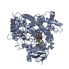
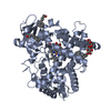
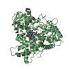
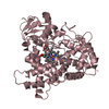
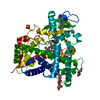
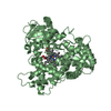
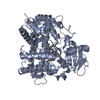
| Title | Crystal Structure of human Cytochrome P450 2B6 (Y226H/K262R) in complex with an analog of a drug Efavirenz | ||||||
 Components Components | Cytochrome P450 2B6 | ||||||
 Keywords Keywords | OXIDOREDUCTASE | ||||||
| Function / homology |  Function and homology information Function and homology informationtestosterone 16-alpha-hydroxylase activity / testosterone 16-beta-hydroxylase activity / Fatty acids / Oxidoreductases; Acting on paired donors, with incorporation or reduction of molecular oxygen; With NADH or NADPH as one donor, and incorporation of one atom of oxygen into the other donor / CYP2E1 reactions / arachidonate epoxygenase activity / ketone metabolic process / epoxygenase P450 pathway / anandamide 8,9 epoxidase activity / anandamide 11,12 epoxidase activity ...testosterone 16-alpha-hydroxylase activity / testosterone 16-beta-hydroxylase activity / Fatty acids / Oxidoreductases; Acting on paired donors, with incorporation or reduction of molecular oxygen; With NADH or NADPH as one donor, and incorporation of one atom of oxygen into the other donor / CYP2E1 reactions / arachidonate epoxygenase activity / ketone metabolic process / epoxygenase P450 pathway / anandamide 8,9 epoxidase activity / anandamide 11,12 epoxidase activity / anandamide 14,15 epoxidase activity / estrogen 2-hydroxylase activity / Xenobiotics / Phase I - Functionalization of compounds / oxidoreductase activity, acting on paired donors, with incorporation or reduction of molecular oxygen, reduced flavin or flavoprotein as one donor, and incorporation of one atom of oxygen / steroid metabolic process / xenobiotic catabolic process / xenobiotic metabolic process / monooxygenase activity / iron ion binding / heme binding / endoplasmic reticulum membrane / cytoplasm Similarity search - Function | ||||||
| Biological species |  Homo sapiens (human) Homo sapiens (human) | ||||||
| Method |  X-RAY DIFFRACTION / SYNCHROTRON / MOLECULAR REPLACEMENT / Resolution: 2.99 Å X-RAY DIFFRACTION / SYNCHROTRON / MOLECULAR REPLACEMENT / Resolution: 2.99 Å | ||||||
 Authors Authors | Shah, M.B. / Halpert, J.R. | ||||||
| Funding support |  United States, 1items United States, 1items
| ||||||
 Citation Citation | Journal: Int J Mol Sci / Year: 2018 Title: Crystal Structure of CYP2B6 in Complex with an Efavirenz Analog. Authors: Shah, M.B. / Zhang, Q. / Halpert, J.R. | ||||||
| History |
|
- Structure visualization
| Structure viewer | Molecule: MolmilJmol/JSmol |
|---|
- Downloads & links
-Download
| PDBx/mmCIF format | 5wbg.cif.gz | 558.8 KB | Display | PDBx/mmCIF format |
|---|---|---|---|---|
| PDB format | pdb5wbg.ent.gz | 458 KB | Display | PDB format |
| PDBx/mmJSON format | 5wbg.json.gz | Tree view | PDBx/mmJSON format | |
| Others |  Other downloads Other downloads |
-Validation report
| Arichive directory | https://data.pdbj.org/pub/pdb/validation_reports/wb/5wbgftp://data.pdbj.org/pub/pdb/validation_reports/wb/5wbg | HTTPS FTP |
|---|
-Related structure data
| Related structure data |  4i91S S: Starting model for refinement |
|---|---|
| Similar structure data |


-Links
 PDBj
PDBj

- Assembly
| Deposited unit | 
| |||||||||||||||||||||||||||||||||||||||||||||||||||||||||||||||||||||||||||||||||||||||||||||||||||||||||||||||||||||||||||||||||||||||||||||||||||||||
|---|---|---|---|---|---|---|---|---|---|---|---|---|---|---|---|---|---|---|---|---|---|---|---|---|---|---|---|---|---|---|---|---|---|---|---|---|---|---|---|---|---|---|---|---|---|---|---|---|---|---|---|---|---|---|---|---|---|---|---|---|---|---|---|---|---|---|---|---|---|---|---|---|---|---|---|---|---|---|---|---|---|---|---|---|---|---|---|---|---|---|---|---|---|---|---|---|---|---|---|---|---|---|---|---|---|---|---|---|---|---|---|---|---|---|---|---|---|---|---|---|---|---|---|---|---|---|---|---|---|---|---|---|---|---|---|---|---|---|---|---|---|---|---|---|---|---|---|---|---|---|---|---|
| 1 | 
| |||||||||||||||||||||||||||||||||||||||||||||||||||||||||||||||||||||||||||||||||||||||||||||||||||||||||||||||||||||||||||||||||||||||||||||||||||||||
| 2 | 
| |||||||||||||||||||||||||||||||||||||||||||||||||||||||||||||||||||||||||||||||||||||||||||||||||||||||||||||||||||||||||||||||||||||||||||||||||||||||
| 3 | 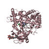
| |||||||||||||||||||||||||||||||||||||||||||||||||||||||||||||||||||||||||||||||||||||||||||||||||||||||||||||||||||||||||||||||||||||||||||||||||||||||
| 4 | 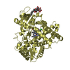
| |||||||||||||||||||||||||||||||||||||||||||||||||||||||||||||||||||||||||||||||||||||||||||||||||||||||||||||||||||||||||||||||||||||||||||||||||||||||
| 5 | 
| |||||||||||||||||||||||||||||||||||||||||||||||||||||||||||||||||||||||||||||||||||||||||||||||||||||||||||||||||||||||||||||||||||||||||||||||||||||||
| 6 | 
| |||||||||||||||||||||||||||||||||||||||||||||||||||||||||||||||||||||||||||||||||||||||||||||||||||||||||||||||||||||||||||||||||||||||||||||||||||||||
| Unit cell |
| |||||||||||||||||||||||||||||||||||||||||||||||||||||||||||||||||||||||||||||||||||||||||||||||||||||||||||||||||||||||||||||||||||||||||||||||||||||||
| Noncrystallographic symmetry (NCS) | NCS domain:
NCS domain segments: Ens-ID: 1 / Refine code: 2
|
-Components
-Protein , 1 types, 6 molecules ABCDEF
| #1: Protein | Mass: 54646.004 Da / Num. of mol.: 6 / Fragment: residues 30-491 / Mutation: Y226H, K262R Source method: isolated from a genetically manipulated source Source: (gene. exp.) Homo sapiens (human) / Gene: CYP2B6 / Plasmid: pKKdHDetails (production host): N-terminally truncated and engineered construct with His-tag at the C-terminus Production host:  References: UniProt: P20813, Oxidoreductases; Acting on paired donors, with incorporation or reduction of molecular oxygen; With NADH or NADPH as one donor, and incorporation of one atom of oxygen into the other donor |
|---|
-Non-polymers , 5 types, 173 molecules 


| #2: Chemical | ChemComp-HEM /  Mass: 616.487 Da / Num. of mol.: 6 / Source method: obtained synthetically / Formula: C34H32FeN4O4 Mass: 616.487 Da / Num. of mol.: 6 / Source method: obtained synthetically / Formula: C34H32FeN4O4#3: Chemical | ChemComp-9ZJ / (  Mass: 315.718 Da / Num. of mol.: 6 / Source method: obtained synthetically / Formula: C15H13ClF3NO Mass: 315.718 Da / Num. of mol.: 6 / Source method: obtained synthetically / Formula: C15H13ClF3NO#4: Chemical | ChemComp-ZAZ / |  Mass: 170.292 Da / Num. of mol.: 1 / Source method: obtained synthetically / Formula: C11H22O Mass: 170.292 Da / Num. of mol.: 1 / Source method: obtained synthetically / Formula: C11H22O#5: Chemical | ChemComp-CM5 /  Mass: 494.573 Da / Num. of mol.: 5 / Source method: obtained synthetically / Formula: C23H42O11 / Comment: detergent*YM Mass: 494.573 Da / Num. of mol.: 5 / Source method: obtained synthetically / Formula: C23H42O11 / Comment: detergent*YM#6: Water | ChemComp-HOH / | Mass: 18.015 Da / Num. of mol.: 155 / Source method: isolated from a natural source / Formula: H2O |
|---|
-Experimental details
-Experiment
| Experiment | Method: X-RAY DIFFRACTION / Number of used crystals: 1 |
|---|
- Sample preparation
| Crystal | Density Matthews: 3.77 Å3/Da / Density % sol: 67.4 % |
|---|---|
| Crystal grow | Temperature: 291 K / Method: vapor diffusion, sitting drop / pH: 7.4 Details: 0.8M Sodium Formate 0.1 M Tris pH 7.5 8% w/v PEG 20000 8% v/v PEG 550 MME |
-Data collection
| Diffraction | Mean temperature: 100 K |
|---|---|
| Diffraction source | Source: SYNCHROTRON / Site: SSRL / Beamline: BL14-1 / Wavelength: 1.1808 |
| Detector | Type: MARMOSAIC 325 mm CCD / Detector: CCD / Date: Jan 8, 2017 |
| Radiation | Monochromator: Si(111) / Protocol: SINGLE WAVELENGTH / Monochromatic (M) / Laue (L): M / Scattering type: x-ray |
| Radiation wavelength | Wavelength: 1.1808 Å / Relative weight: 1 |
| Reflection | Resolution: 2.99→50 Å / Num. obs: 89926 / % possible obs: 98.9 % / Redundancy: 3.3 % / Rmerge(I) obs: 0.188 / Net I/σ(I): 6.4 |
| Reflection shell | Resolution: 2.99→3.07 Å / Redundancy: 3.2 % / Rmerge(I) obs: 0.1 / Mean I/σ(I) obs: 1.6 / % possible all: 96.7 |
- Processing
| Software |
| ||||||||||||||||||||||||||||||||||||||||||||||||||||||||||||||||||||||||||||||||||||||||||||||||||||||||||||||||||||||||||||||||||||||||||||||||||||||||||||||||||||||||||||||||||||||
|---|---|---|---|---|---|---|---|---|---|---|---|---|---|---|---|---|---|---|---|---|---|---|---|---|---|---|---|---|---|---|---|---|---|---|---|---|---|---|---|---|---|---|---|---|---|---|---|---|---|---|---|---|---|---|---|---|---|---|---|---|---|---|---|---|---|---|---|---|---|---|---|---|---|---|---|---|---|---|---|---|---|---|---|---|---|---|---|---|---|---|---|---|---|---|---|---|---|---|---|---|---|---|---|---|---|---|---|---|---|---|---|---|---|---|---|---|---|---|---|---|---|---|---|---|---|---|---|---|---|---|---|---|---|---|---|---|---|---|---|---|---|---|---|---|---|---|---|---|---|---|---|---|---|---|---|---|---|---|---|---|---|---|---|---|---|---|---|---|---|---|---|---|---|---|---|---|---|---|---|---|---|---|---|
| Refinement | Method to determine structure: MOLECULAR REPLACEMENT Starting model: 4I91 Resolution: 2.99→50 Å / Cor.coef. Fo:Fc: 0.922 / Cor.coef. Fo:Fc free: 0.896 / SU B: 20.491 / SU ML: 0.37 / Cross valid method: THROUGHOUT / ESU R: 0.699 / ESU R Free: 0.4 / Details: HYDROGENS HAVE BEEN ADDED IN THE RIDING POSITIONS
| ||||||||||||||||||||||||||||||||||||||||||||||||||||||||||||||||||||||||||||||||||||||||||||||||||||||||||||||||||||||||||||||||||||||||||||||||||||||||||||||||||||||||||||||||||||||
| Solvent computation | Ion probe radii: 0.8 Å / Shrinkage radii: 0.8 Å / VDW probe radii: 1.4 Å | ||||||||||||||||||||||||||||||||||||||||||||||||||||||||||||||||||||||||||||||||||||||||||||||||||||||||||||||||||||||||||||||||||||||||||||||||||||||||||||||||||||||||||||||||||||||
| Displacement parameters | Biso mean: 49.63 Å2
| ||||||||||||||||||||||||||||||||||||||||||||||||||||||||||||||||||||||||||||||||||||||||||||||||||||||||||||||||||||||||||||||||||||||||||||||||||||||||||||||||||||||||||||||||||||||
| Refinement step | Cycle: LAST / Resolution: 2.99→50 Å
| ||||||||||||||||||||||||||||||||||||||||||||||||||||||||||||||||||||||||||||||||||||||||||||||||||||||||||||||||||||||||||||||||||||||||||||||||||||||||||||||||||||||||||||||||||||||
| Refine LS restraints |
|