 Movie
Movie Controller

[English] 日本語
 Yorodumi
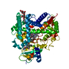
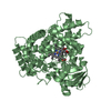
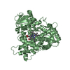
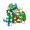
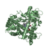
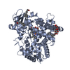
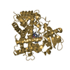
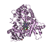
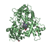
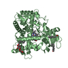
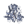
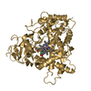
- PDB-3lc4: Human Cytochrome P450 2E1 in Complex with Omega-Imidazolyl-Dodeca... -
+ Open data

- Basic information
| Entry | Database: PDB / ID: 3lc4 | ||||||
|---|---|---|---|---|---|---|---|
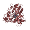
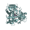
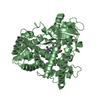
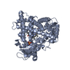
| Title | Human Cytochrome P450 2E1 in Complex with Omega-Imidazolyl-Dodecanoic Acid | ||||||
 Components Components | Cytochrome P450 2E1 | ||||||
 Keywords Keywords | OXIDOREDUCTASE / CYP2E1 / P450 2E1 / monooxygenase / acetaminophen / heme / endoplasmic reticulum / iron / membrane / metal-binding / microsome | ||||||
| Function / homology |  Function and homology information Function and homology information: / carbon tetrachloride metabolic process / benzene metabolic process / 4-nitrophenol metabolic process / halogenated hydrocarbon metabolic process / 4-nitrophenol 2-monooxygenase activity / long-chain fatty acid omega-1 hydroxylase activity / CYP2E1 reactions / long-chain fatty acid metabolic process / arachidonate epoxygenase activity ...: / carbon tetrachloride metabolic process / benzene metabolic process / 4-nitrophenol metabolic process / halogenated hydrocarbon metabolic process / 4-nitrophenol 2-monooxygenase activity / long-chain fatty acid omega-1 hydroxylase activity / CYP2E1 reactions / long-chain fatty acid metabolic process / arachidonate epoxygenase activity / epoxygenase P450 pathway / lipid hydroxylation / Biosynthesis of maresin-like SPMs / oxidoreductase activity, acting on paired donors, with incorporation or reduction of molecular oxygen, NAD(P)H as one donor, and incorporation of one atom of oxygen / monoterpenoid metabolic process / Xenobiotics / Paracetamol ADME / oxidoreductase activity, acting on paired donors, with incorporation or reduction of molecular oxygen, reduced flavin or flavoprotein as one donor, and incorporation of one atom of oxygen / unspecific monooxygenase / long-chain fatty acid biosynthetic process / Aspirin ADME / steroid metabolic process / xenobiotic metabolic process / response to bacterium / Hsp70 protein binding / monooxygenase activity / oxygen binding / Hsp90 protein binding / oxidoreductase activity / mitochondrial inner membrane / iron ion binding / heme binding / endoplasmic reticulum membrane / enzyme binding / cytoplasm Similarity search - Function | ||||||
| Biological species |  Homo sapiens (human) Homo sapiens (human) | ||||||
| Method |  X-RAY DIFFRACTION / SYNCHROTRON / MOLECULAR REPLACEMENT / Resolution: 3.1 Å X-RAY DIFFRACTION / SYNCHROTRON / MOLECULAR REPLACEMENT / Resolution: 3.1 Å | ||||||
 Authors Authors | Scott, E.E. / Porubsky, P.R. | ||||||
 Citation Citation | Journal: J.Biol.Chem. / Year: 2010 Title: Human cytochrome P450 2E1 structures with fatty acid analogs reveal a previously unobserved binding mode. Authors: Porubsky, P.R. / Battaile, K.P. / Scott, E.E. | ||||||
| History |
|
- Structure visualization
| Structure viewer | Molecule: MolmilJmol/JSmol |
|---|
- Downloads & links
-Download
| PDBx/mmCIF format | 3lc4.cif.gz | 197.7 KB | Display | PDBx/mmCIF format |
|---|---|---|---|---|
| PDB format | pdb3lc4.ent.gz | 156.8 KB | Display | PDB format |
| PDBx/mmJSON format | 3lc4.json.gz | Tree view | PDBx/mmJSON format | |
| Others |  Other downloads Other downloads |
-Validation report
| Arichive directory | https://data.pdbj.org/pub/pdb/validation_reports/lc/3lc4ftp://data.pdbj.org/pub/pdb/validation_reports/lc/3lc4 | HTTPS FTP |
|---|
-Related structure data
| Related structure data |  3gphC  3kohC  3e6iS C: citing same article ( S: Starting model for refinement |
|---|---|
| Similar structure data |


-Links
 PDBj
PDBj


- Assembly
| Deposited unit | 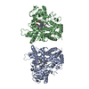
| ||||||||
|---|---|---|---|---|---|---|---|---|---|
| 1 | 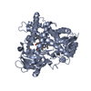
| ||||||||
| 2 | 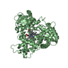
| ||||||||
| Unit cell |
|
-Components
| #1: Protein | Mass: 55058.488 Da / Num. of mol.: 2 / Fragment: sequence database residues 32-493 Source method: isolated from a genetically manipulated source Source: (gene. exp.) Homo sapiens (human) / Gene: CYP2E, CYP2E1 / Plasmid: pKK2E1dH / Production host:  References: UniProt: P05181, Oxidoreductases; Acting on paired donors, with incorporation or reduction of molecular oxygen; With NADH or NADPH as one donor, and incorporation of one atom of oxygen into the other donor #2: Chemical |   Mass: 616.487 Da / Num. of mol.: 2 / Source method: obtained synthetically / Formula: C34H32FeN4O4 Mass: 616.487 Da / Num. of mol.: 2 / Source method: obtained synthetically / Formula: C34H32FeN4O4#3: Chemical | 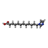  Mass: 266.379 Da / Num. of mol.: 2 / Source method: obtained synthetically / Formula: C15H26N2O2 Mass: 266.379 Da / Num. of mol.: 2 / Source method: obtained synthetically / Formula: C15H26N2O2#4: Water | ChemComp-HOH / |  Mass: 18.015 Da / Num. of mol.: 5 / Source method: isolated from a natural source / Formula: H2O Mass: 18.015 Da / Num. of mol.: 5 / Source method: isolated from a natural source / Formula: H2O |
|---|
-Experimental details
-Experiment
| Experiment | Method: X-RAY DIFFRACTION / Number of used crystals: 1 |
|---|
- Sample preparation
| Crystal | Density Matthews: 2.55 Å3/Da / Density % sol: 51.86 % |
|---|---|
| Crystal grow | Temperature: 293 K / Method: vapor diffusion, hanging drop / pH: 7.5 Details: NaHEPES pH 7.5, 5% iso-propanol, 22% PEG 2000 MME, VAPOR DIFFUSION, HANGING DROP, temperature 293K |
-Data collection
| Diffraction | Mean temperature: 100 K |
|---|---|
| Diffraction source | Source: SYNCHROTRON / Site: SSRL  / Beamline: BL9-2 / Wavelength: 0.98 Å / Beamline: BL9-2 / Wavelength: 0.98 Å |
| Detector | Type: ADSC QUANTUM 315 / Detector: CCD / Date: May 1, 2009 / Details: mirrors |
| Radiation | Monochromator: SIDE-SCATTERING CUBEROOT I- BEAM BENT SINGLE CRYSTAL. ASYMMETRIC CUT 12.2 DEGS Protocol: SINGLE WAVELENGTH / Monochromatic (M) / Laue (L): M / Scattering type: x-ray |
| Radiation wavelength | Wavelength: 0.98 Å / Relative weight: 1 |
| Reflection | Resolution: 3.1→38.01 Å / Num. obs: 19925 / % possible obs: 100 % / Redundancy: 12.3 % / Rsym value: 0.189 |
| Reflection shell | Resolution: 3.1→3.18 Å / Redundancy: 7.4 % / Mean I/σ(I) obs: 1.7 / Num. unique all: 1443 / Rsym value: 0.405 / % possible all: 100 |
- Processing
| Software |
| ||||||||||||||||||||||||||||||||||||||||||||||||||||||||||||||||||||||||||||||||||||||||||||||||||||||||||||||||||||||||||||||||||||||||||||||||||||||||||||||||||||||||||
|---|---|---|---|---|---|---|---|---|---|---|---|---|---|---|---|---|---|---|---|---|---|---|---|---|---|---|---|---|---|---|---|---|---|---|---|---|---|---|---|---|---|---|---|---|---|---|---|---|---|---|---|---|---|---|---|---|---|---|---|---|---|---|---|---|---|---|---|---|---|---|---|---|---|---|---|---|---|---|---|---|---|---|---|---|---|---|---|---|---|---|---|---|---|---|---|---|---|---|---|---|---|---|---|---|---|---|---|---|---|---|---|---|---|---|---|---|---|---|---|---|---|---|---|---|---|---|---|---|---|---|---|---|---|---|---|---|---|---|---|---|---|---|---|---|---|---|---|---|---|---|---|---|---|---|---|---|---|---|---|---|---|---|---|---|---|---|---|---|---|---|---|
| Refinement | Method to determine structure: MOLECULAR REPLACEMENT Starting model: PDB entry 3E6I Resolution: 3.1→37.93 Å / Cor.coef. Fo:Fc: 0.918 / Cor.coef. Fo:Fc free: 0.848 / SU B: 24.186 / SU ML: 0.429 / Cross valid method: THROUGHOUT / ESU R Free: 0.543 / Stereochemistry target values: MAXIMUM LIKELIHOOD
| ||||||||||||||||||||||||||||||||||||||||||||||||||||||||||||||||||||||||||||||||||||||||||||||||||||||||||||||||||||||||||||||||||||||||||||||||||||||||||||||||||||||||||
| Solvent computation | Ion probe radii: 0.8 Å / Shrinkage radii: 0.8 Å / VDW probe radii: 1.4 Å / Solvent model: BABINET MODEL WITH MASK | ||||||||||||||||||||||||||||||||||||||||||||||||||||||||||||||||||||||||||||||||||||||||||||||||||||||||||||||||||||||||||||||||||||||||||||||||||||||||||||||||||||||||||
| Displacement parameters | Biso mean: 47.046 Å2
| ||||||||||||||||||||||||||||||||||||||||||||||||||||||||||||||||||||||||||||||||||||||||||||||||||||||||||||||||||||||||||||||||||||||||||||||||||||||||||||||||||||||||||
| Refinement step | Cycle: LAST / Resolution: 3.1→37.93 Å
| ||||||||||||||||||||||||||||||||||||||||||||||||||||||||||||||||||||||||||||||||||||||||||||||||||||||||||||||||||||||||||||||||||||||||||||||||||||||||||||||||||||||||||
| Refine LS restraints |
| ||||||||||||||||||||||||||||||||||||||||||||||||||||||||||||||||||||||||||||||||||||||||||||||||||||||||||||||||||||||||||||||||||||||||||||||||||||||||||||||||||||||||||
| LS refinement shell | Resolution: 3.103→3.183 Å / Total num. of bins used: 20
|
