 Movie
Movie Controller


+ Open data

- Basic information
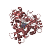
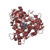
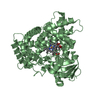
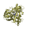
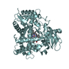
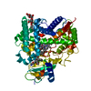
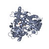
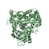
| Entry | Database: PDB / ID: 2pg7 | ||||||
|---|---|---|---|---|---|---|---|
| Title | Crystal Structure of Human Microsomal P450 2A6 N297Q/I300V | ||||||
 Components Components | Cytochrome P450 2A6 | ||||||
 Keywords Keywords | OXIDOREDUCTASE / CYP2A6 / P450 2A6 / P450 / Monooxygenases / drug metabolizing enzyme / heme / indole / mutant | ||||||
| Function / homology |  Function and homology information Function and homology informationcoumarin catabolic process / coumarin 7-hydroxylase activity / Fatty acids / coumarin metabolic process / Oxidoreductases; Acting on paired donors, with incorporation or reduction of molecular oxygen; With reduced flavin or flavoprotein as one donor, and incorporation of one atom of oxygen into the other donor / CYP2E1 reactions / arachidonate epoxygenase activity / epoxygenase P450 pathway / aflatoxin metabolic process / Aflatoxin activation and detoxification ...coumarin catabolic process / coumarin 7-hydroxylase activity / Fatty acids / coumarin metabolic process / Oxidoreductases; Acting on paired donors, with incorporation or reduction of molecular oxygen; With reduced flavin or flavoprotein as one donor, and incorporation of one atom of oxygen into the other donor / CYP2E1 reactions / arachidonate epoxygenase activity / epoxygenase P450 pathway / aflatoxin metabolic process / Aflatoxin activation and detoxification / Xenobiotics / oxidoreductase activity, acting on paired donors, with incorporation or reduction of molecular oxygen, reduced flavin or flavoprotein as one donor, and incorporation of one atom of oxygen / unspecific monooxygenase / steroid metabolic process / xenobiotic catabolic process / cytoplasmic microtubule / xenobiotic metabolic process / monooxygenase activity / iron ion binding / heme binding / endoplasmic reticulum membrane / enzyme binding / cytoplasm Similarity search - Function | ||||||
| Biological species |  Homo sapiens (human) Homo sapiens (human) | ||||||
| Method |  X-RAY DIFFRACTION / SYNCHROTRON / MOLECULAR REPLACEMENT / Resolution: 2.8 Å X-RAY DIFFRACTION / SYNCHROTRON / MOLECULAR REPLACEMENT / Resolution: 2.8 Å | ||||||
 Authors Authors | Sansen, S. / Hsu, M.H. / Stout, C.D. / Johnson, E.F. | ||||||
 Citation Citation | Journal: Arch.Biochem.Biophys. / Year: 2007 Title: Structural insight into the altered substrate specificity of human cytochrome P450 2A6 mutants. Authors: Sansen, S. / Hsu, M.H. / Stout, C.D. / Johnson, E.F. | ||||||
| History |
|
- Structure visualization
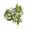
| Structure viewer | Molecule: MolmilJmol/JSmol |
|---|
- Downloads & links
-Download
| PDBx/mmCIF format | 2pg7.cif.gz | 342.2 KB | Display | PDBx/mmCIF format |
|---|---|---|---|---|
| PDB format | pdb2pg7.ent.gz | 285.8 KB | Display | PDB format |
| PDBx/mmJSON format | 2pg7.json.gz | Tree view | PDBx/mmJSON format | |
| Others |  Other downloads Other downloads |
-Validation report
| Arichive directory | https://data.pdbj.org/pub/pdb/validation_reports/pg/2pg7ftp://data.pdbj.org/pub/pdb/validation_reports/pg/2pg7 | HTTPS FTP |
|---|
-Related structure data
| Related structure data |  2pg5C  2pg6C  1z10S S: Starting model for refinement C: citing same article ( |
|---|---|
| Similar structure data |


-Links
 PDBj
PDBj


- Assembly
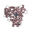
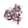
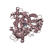
| Deposited unit | 
| ||||||||
|---|---|---|---|---|---|---|---|---|---|
| 1 | 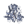
| ||||||||
| 2 | 
| ||||||||
| 3 | 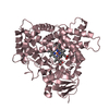
| ||||||||
| 4 | 
| ||||||||
| Unit cell |
| ||||||||
| Details | The biological assembly has not been determined but thought to be a momoner |
-Components
| #1: Protein | Mass: 54671.637 Da / Num. of mol.: 4 / Mutation: N297Q, I300V Source method: isolated from a genetically manipulated source Source: (gene. exp.) Homo sapiens (human) / Gene: CYP2A6 / Plasmid: PCWORI / Production host:  References: UniProt: P11509, UniProt: Q16696*PLUS, unspecific monooxygenase #2: Chemical | ChemComp-HEM /   Mass: 616.487 Da / Num. of mol.: 4 / Source method: obtained synthetically / Formula: C34H32FeN4O4 Mass: 616.487 Da / Num. of mol.: 4 / Source method: obtained synthetically / Formula: C34H32FeN4O4#3: Water | ChemComp-HOH / |  Mass: 18.015 Da / Num. of mol.: 91 / Source method: isolated from a natural source / Formula: H2O Mass: 18.015 Da / Num. of mol.: 91 / Source method: isolated from a natural source / Formula: H2O |
|---|
-Experimental details
-Experiment
| Experiment | Method: X-RAY DIFFRACTION / Number of used crystals: 1 |
|---|
- Sample preparation
| Crystal | Density Matthews: 2.69 Å3/Da / Density % sol: 54.21 % |
|---|---|
| Crystal grow | Temperature: 291 K / Method: vapor diffusion, sitting drop / pH: 8.5 Details: PEG3350, Tris, Ammonium sulfate, Anapoe-35, pH 8.5, VAPOR DIFFUSION, SITTING DROP, temperature 291K |
-Data collection
| Diffraction | Mean temperature: 100 K |
|---|---|
| Diffraction source | Source: SYNCHROTRON / Site: SSRL  / Beamline: BL9-1 / Wavelength: 0.98 Å / Beamline: BL9-1 / Wavelength: 0.98 Å |
| Detector | Type: ADSC QUANTUM 315 / Detector: CCD / Date: Jun 19, 2006 Details: Vertical focussing mirror, singe crystal Si(311) bent monochromator (horizontal focussing) |
| Radiation | Monochromator: Side-scattering cuberoot I-beam bent crystal; asymetric cut 12.2 degs. Protocol: SINGLE WAVELENGTH / Monochromatic (M) / Laue (L): M / Scattering type: x-ray |
| Radiation wavelength | Wavelength: 0.98 Å / Relative weight: 1 |
| Reflection | Resolution: 2.8→104.257 Å / Num. all: 56574 / Num. obs: 56574 / % possible obs: 99.6 % / Observed criterion σ(F): 0 / Observed criterion σ(I): 0 / Redundancy: 4.8 % / Biso Wilson estimate: 69.156 Å2 / Rmerge(I) obs: 0.13 / Rsym value: 0.13 / Net I/σ(I): 4.5 |
| Reflection shell | Resolution: 2.8→2.87 Å / Redundancy: 4.6 % / Rmerge(I) obs: 0.63 / Mean I/σ(I) obs: 1.2 / Num. measured all: 19268 / Num. unique all: 4147 / Rsym value: 0.63 / % possible all: 99.4 |
- Processing
| Software |
| ||||||||||||||||||||||||||||||||
|---|---|---|---|---|---|---|---|---|---|---|---|---|---|---|---|---|---|---|---|---|---|---|---|---|---|---|---|---|---|---|---|---|---|
| Refinement | Method to determine structure: MOLECULAR REPLACEMENT Starting model: coumarin complex of CYP2A6, PDB entry 1Z10 Resolution: 2.8→40 Å / Isotropic thermal model: restrained / Cross valid method: THROUGHOUT / σ(F): 0 / σ(I): 0 / Stereochemistry target values: Engh & Huber
| ||||||||||||||||||||||||||||||||
| Solvent computation | Bsol: 10 Å2 | ||||||||||||||||||||||||||||||||
| Displacement parameters | Biso mean: 53.262 Å2
| ||||||||||||||||||||||||||||||||
| Refine analyze |
| ||||||||||||||||||||||||||||||||
| Refinement step | Cycle: LAST / Resolution: 2.8→40 Å
| ||||||||||||||||||||||||||||||||
| LS refinement shell | Resolution: 2.8→2.93 Å / Rfactor Rfree error: 0.022
| ||||||||||||||||||||||||||||||||
| Xplor file |
|
