 Movie
Movie Controller

[English] 日本語
 Yorodumi
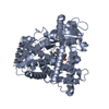
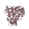
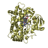
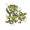
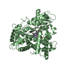
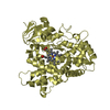
- PDB-1z11: Crystal Structure of Human Microsomal P450 2A6 with Methoxsalen Bound -
+ Open data

- Basic information
| Entry | Database: PDB / ID: 1z11 | ||||||
|---|---|---|---|---|---|---|---|
| Title | Crystal Structure of Human Microsomal P450 2A6 with Methoxsalen Bound | ||||||
 Components Components | cytochrome P450, family 2, subfamily A, polypeptide 6 | ||||||
 Keywords Keywords | OXIDOREDUCTASE / CYP2A6 / P450 2A6 / P450 / monooxygenase / drug metabolizing enzyme / coumarin 7-hydroxylase / nicotine oxidase / heme / methoxsalen | ||||||
| Function / homology |  Function and homology information Function and homology informationcoumarin catabolic process / coumarin 7-hydroxylase activity / coumarin metabolic process / Oxidoreductases; Acting on paired donors, with incorporation or reduction of molecular oxygen; With reduced flavin or flavoprotein as one donor, and incorporation of one atom of oxygen into the other donor / CYP2E1 reactions / arachidonate epoxygenase activity / epoxygenase P450 pathway / Xenobiotics / oxidoreductase activity, acting on paired donors, with incorporation or reduction of molecular oxygen, reduced flavin or flavoprotein as one donor, and incorporation of one atom of oxygen / steroid metabolic process ...coumarin catabolic process / coumarin 7-hydroxylase activity / coumarin metabolic process / Oxidoreductases; Acting on paired donors, with incorporation or reduction of molecular oxygen; With reduced flavin or flavoprotein as one donor, and incorporation of one atom of oxygen into the other donor / CYP2E1 reactions / arachidonate epoxygenase activity / epoxygenase P450 pathway / Xenobiotics / oxidoreductase activity, acting on paired donors, with incorporation or reduction of molecular oxygen, reduced flavin or flavoprotein as one donor, and incorporation of one atom of oxygen / steroid metabolic process / xenobiotic catabolic process / cytoplasmic microtubule / xenobiotic metabolic process / iron ion binding / heme binding / endoplasmic reticulum membrane / enzyme binding / cytoplasm Similarity search - Function | ||||||
| Biological species |  Homo sapiens (human) Homo sapiens (human) | ||||||
| Method |  X-RAY DIFFRACTION / SYNCHROTRON / MOLECULAR REPLACEMENT / Resolution: 2.05 Å X-RAY DIFFRACTION / SYNCHROTRON / MOLECULAR REPLACEMENT / Resolution: 2.05 Å | ||||||
 Authors Authors | Yano, J.K. / Hsu, M.H. / Griffin, K.J. / Stout, C.D. / Johnson, E.F. | ||||||
 Citation Citation | Journal: Nat.Struct.Mol.Biol. / Year: 2005 Title: Structures of human microsomal cytochrome P450 2A6 complexed with coumarin and methoxsalen Authors: Yano, J.K. / Hsu, M.H. / Griffin, K.J. / Stout, C.D. / Johnson, E.F. | ||||||
| History |
| ||||||
| Remark 999 | SEQUENCE Residues 1-28 were replaced with the sequence MAKKTS. |
- Structure visualization
| Structure viewer | Molecule: MolmilJmol/JSmol |
|---|
- Downloads & links
-Download
| PDBx/mmCIF format | 1z11.cif.gz | 387.6 KB | Display | PDBx/mmCIF format |
|---|---|---|---|---|
| PDB format | pdb1z11.ent.gz | 318.3 KB | Display | PDB format |
| PDBx/mmJSON format | 1z11.json.gz | Tree view | PDBx/mmJSON format | |
| Others |  Other downloads Other downloads |
-Validation report
| Arichive directory | https://data.pdbj.org/pub/pdb/validation_reports/z1/1z11ftp://data.pdbj.org/pub/pdb/validation_reports/z1/1z11 | HTTPS FTP |
|---|
-Related structure data
| Related structure data |  1z10SC S: Starting model for refinement C: citing same article ( |
|---|---|
| Similar structure data |


-Links
 PDBj
PDBj

- Assembly
| Deposited unit | 
| ||||||||
|---|---|---|---|---|---|---|---|---|---|
| 1 | 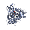
| ||||||||
| 2 | 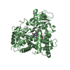
| ||||||||
| 3 | 
| ||||||||
| 4 | 
| ||||||||
| Unit cell |
| ||||||||
| Details | The biological assembly has not been determined but thought to be a monomer. |
-Components
| #1: Protein | Mass: 54671.637 Da / Num. of mol.: 4 / Fragment: catalytic domain Source method: isolated from a genetically manipulated source Source: (gene. exp.) Homo sapiens (human) / Gene: CYP2A6 / Plasmid: pCWori / Production host:  #2: Chemical | ChemComp-HEM /   Mass: 616.487 Da / Num. of mol.: 4 / Source method: obtained synthetically / Formula: C34H32FeN4O4 Mass: 616.487 Da / Num. of mol.: 4 / Source method: obtained synthetically / Formula: C34H32FeN4O4#3: Chemical | ChemComp-8MO / 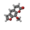  Mass: 216.190 Da / Num. of mol.: 4 / Source method: obtained synthetically / Formula: C12H8O4 Mass: 216.190 Da / Num. of mol.: 4 / Source method: obtained synthetically / Formula: C12H8O4#4: Water | ChemComp-HOH / |  Mass: 18.015 Da / Num. of mol.: 504 / Source method: isolated from a natural source / Formula: H2O Mass: 18.015 Da / Num. of mol.: 504 / Source method: isolated from a natural source / Formula: H2O |
|---|
-Experimental details
-Experiment
| Experiment | Method: X-RAY DIFFRACTION / Number of used crystals: 1 |
|---|
- Sample preparation
| Crystal | Density Matthews: 2.7 Å3/Da / Density % sol: 51.8 % |
|---|---|
| Crystal grow | Temperature: 291 K / Method: vapor diffusion, sitting drop / pH: 8.5 Details: PEG3350, Tris, ammonium sulfate, Anapoe-X-405, pH 8.5, VAPOR DIFFUSION, SITTING DROP, temperature 291K |
-Data collection
| Diffraction | Mean temperature: 100 K | |||||||||
|---|---|---|---|---|---|---|---|---|---|---|
| Diffraction source | Source: SYNCHROTRON / Site: SSRL  / Beamline: BL9-2 / Wavelength: 1.03, 0.98 / Beamline: BL9-2 / Wavelength: 1.03, 0.98 | |||||||||
| Detector | Type: ADSC QUANTUM 315 / Detector: CCD / Date: Apr 4, 2004 / Details: double crystal monochromator | |||||||||
| Radiation | Protocol: SINGLE WAVELENGTH / Monochromatic (M) / Laue (L): M / Scattering type: x-ray | |||||||||
| Radiation wavelength |
| |||||||||
| Reflection | Resolution: 2.05→50 Å / Num. all: 143181 / Num. obs: 140326 / % possible obs: 98 % / Observed criterion σ(F): 0 / Observed criterion σ(I): 0 / Redundancy: 2.5 % / Rmerge(I) obs: 0.046 / Net I/σ(I): 30 | |||||||||
| Reflection shell | Resolution: 2.05→2.12 Å / Redundancy: 2.4 % / Rmerge(I) obs: 0.243 / Mean I/σ(I) obs: 2.6 / Num. unique all: 12584 / % possible all: 88.9 |
- Processing
| Software |
| |||||||||||||||||||||||||
|---|---|---|---|---|---|---|---|---|---|---|---|---|---|---|---|---|---|---|---|---|---|---|---|---|---|---|
| Refinement | Method to determine structure: MOLECULAR REPLACEMENT Starting model: Coumarin complex of CYP2A6, pdb entry 1Z10 Resolution: 2.05→50 Å / Cross valid method: THROUGHOUT / σ(F): 0 / σ(I): 0 / Stereochemistry target values: Engh & Huber
| |||||||||||||||||||||||||
| Refine analyze |
| |||||||||||||||||||||||||
| Refinement step | Cycle: LAST / Resolution: 2.05→50 Å
| |||||||||||||||||||||||||
| Refine LS restraints |
|
