 Movie
Movie Controller


+ Open data

- Basic information
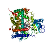
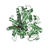
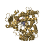
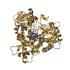
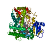
| Entry | Database: PDB / ID: 1pq2 | ||||||
|---|---|---|---|---|---|---|---|
| Title | Crystal Structure of Human Drug Metabolizing Cytochrome P450 2C8 | ||||||
 Components Components | Cytochrome P450 2C8 | ||||||
 Keywords Keywords | OXIDOREDUCTASE / CYTOCHROME P450 / CYP2C8 / MEMBRANE PROTEIN / TAXOL 6-HYDROXYLASE | ||||||
| Function / homology |  Function and homology information Function and homology information: / Synthesis of (16-20)-hydroxyeicosatetraenoic acids (HETE) / omega-hydroxylase P450 pathway / CYP2E1 reactions / arachidonate epoxygenase activity / epoxygenase P450 pathway / icosanoid biosynthetic process / retinoic acid 4-hydroxylase activity / caffeine oxidase activity / estrogen 16-alpha-hydroxylase activity ...: / Synthesis of (16-20)-hydroxyeicosatetraenoic acids (HETE) / omega-hydroxylase P450 pathway / CYP2E1 reactions / arachidonate epoxygenase activity / epoxygenase P450 pathway / icosanoid biosynthetic process / retinoic acid 4-hydroxylase activity / caffeine oxidase activity / estrogen 16-alpha-hydroxylase activity / lipid hydroxylation / Biosynthesis of maresin-like SPMs / Synthesis of epoxy (EET) and dihydroxyeicosatrienoic acids (DHET) / oxidative demethylation / Xenobiotics / oxidoreductase activity, acting on paired donors, with incorporation or reduction of molecular oxygen, reduced flavin or flavoprotein as one donor, and incorporation of one atom of oxygen / estrogen metabolic process / retinoic acid metabolic process / retinol metabolic process / unspecific monooxygenase / long-chain fatty acid biosynthetic process / Aspirin ADME / steroid metabolic process / xenobiotic catabolic process / xenobiotic metabolic process / monooxygenase activity / iron ion binding / heme binding / endoplasmic reticulum membrane / plasma membrane / cytoplasm Similarity search - Function | ||||||
| Biological species |  Homo sapiens (human) Homo sapiens (human) | ||||||
| Method |  X-RAY DIFFRACTION / SYNCHROTRON / MOLECULAR REPLACEMENT / Resolution: 2.7 Å X-RAY DIFFRACTION / SYNCHROTRON / MOLECULAR REPLACEMENT / Resolution: 2.7 Å | ||||||
 Authors Authors | Schoch, G.A. / Yano, J.K. / Wester, M.R. / Griffin, K.J. / Stout, C.D. / Johnson, E.F. | ||||||
 Citation Citation | Journal: J.Biol.Chem. / Year: 2004 Title: Structure of human microsomal cytochrome P450 2C8. Evidence for a peripheral fatty acid binding site Authors: Schoch, G.A. / Yano, J.K. / Wester, M.R. / Griffin, K.J. / Stout, C.D. / Johnson, E.F. | ||||||
| History |
|
- Structure visualization
| Structure viewer | Molecule: MolmilJmol/JSmol |
|---|
- Downloads & links
-Download
| PDBx/mmCIF format | 1pq2.cif.gz | 196.9 KB | Display | PDBx/mmCIF format |
|---|---|---|---|---|
| PDB format | pdb1pq2.ent.gz | 156.4 KB | Display | PDB format |
| PDBx/mmJSON format | 1pq2.json.gz | Tree view | PDBx/mmJSON format | |
| Others |  Other downloads Other downloads |
-Validation report
| Arichive directory | https://data.pdbj.org/pub/pdb/validation_reports/pq/1pq2ftp://data.pdbj.org/pub/pdb/validation_reports/pq/1pq2 | HTTPS FTP |
|---|
-Related structure data
| Related structure data |  1n6bS S: Starting model for refinement |
|---|---|
| Similar structure data |


-Links
 PDBj
PDBj


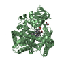
- Assembly
| Deposited unit | 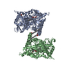
| ||||||||
|---|---|---|---|---|---|---|---|---|---|
| 1 | 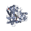
| ||||||||
| 2 | 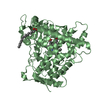
| ||||||||
| Unit cell |
|
-Components
| #1: Protein | Mass: 54054.156 Da / Num. of mol.: 2 Source method: isolated from a genetically manipulated source Source: (gene. exp.) Homo sapiens (human) / Gene: CYP2C8 / Production host:  #2: Chemical | ChemComp-PO4 / |   Mass: 94.971 Da / Num. of mol.: 1 / Source method: obtained synthetically / Formula: PO4 Mass: 94.971 Da / Num. of mol.: 1 / Source method: obtained synthetically / Formula: PO4#3: Chemical |   Mass: 616.487 Da / Num. of mol.: 2 / Source method: obtained synthetically / Formula: C34H32FeN4O4 Mass: 616.487 Da / Num. of mol.: 2 / Source method: obtained synthetically / Formula: C34H32FeN4O4#4: Chemical |   Mass: 256.424 Da / Num. of mol.: 2 / Source method: obtained synthetically / Formula: C16H32O2 Mass: 256.424 Da / Num. of mol.: 2 / Source method: obtained synthetically / Formula: C16H32O2#5: Water | ChemComp-HOH / |  Mass: 18.015 Da / Num. of mol.: 37 / Source method: isolated from a natural source / Formula: H2O Mass: 18.015 Da / Num. of mol.: 37 / Source method: isolated from a natural source / Formula: H2O |
|---|
-Experimental details
-Experiment
| Experiment | Method: X-RAY DIFFRACTION / Number of used crystals: 1 |
|---|
- Sample preparation
| Crystal | Density Matthews: 3.03 Å3/Da / Density % sol: 59.36 % | |||||||||||||||||||||||||||||||||||||||||||||||||||||||||||||||||||||||||||||||||||||||||||
|---|---|---|---|---|---|---|---|---|---|---|---|---|---|---|---|---|---|---|---|---|---|---|---|---|---|---|---|---|---|---|---|---|---|---|---|---|---|---|---|---|---|---|---|---|---|---|---|---|---|---|---|---|---|---|---|---|---|---|---|---|---|---|---|---|---|---|---|---|---|---|---|---|---|---|---|---|---|---|---|---|---|---|---|---|---|---|---|---|---|---|---|---|
| Crystal grow | Temperature: 298 K / Method: vapor diffusion, sitting drop / pH: 7.5 Details: ETHANOL, PEG 4000, HEPES, SODIUM CHLORIDE, CYMAL-6 , pH 7.5, VAPOR DIFFUSION, SITTING DROP, temperature 298K | |||||||||||||||||||||||||||||||||||||||||||||||||||||||||||||||||||||||||||||||||||||||||||
| Crystal grow | *PLUS Temperature: 24 ℃ / Method: vapor diffusion, sitting drop | |||||||||||||||||||||||||||||||||||||||||||||||||||||||||||||||||||||||||||||||||||||||||||
| Components of the solutions | *PLUS
|
-Data collection
| Diffraction | Mean temperature: 100 K |
|---|---|
| Diffraction source | Source: SYNCHROTRON / Site: SSRL  / Beamline: BL7-1 / Wavelength: 1.08 Å / Beamline: BL7-1 / Wavelength: 1.08 Å |
| Detector | Type: MAR scanner 345 mm plate / Detector: IMAGE PLATE / Date: Dec 13, 2002 |
| Radiation | Protocol: SINGLE WAVELENGTH / Monochromatic (M) / Laue (L): M / Scattering type: x-ray |
| Radiation wavelength | Wavelength: 1.08 Å / Relative weight: 1 |
| Reflection | Highest resolution: 2.6 Å / Num. all: 82164 / Num. obs: 38682 / % possible obs: 97.9 % / Observed criterion σ(F): 0 / Observed criterion σ(I): 0 / Redundancy: 2.1 % / Rsym value: 0.052 / Net I/σ(I): 10.9 |
| Reflection shell | Resolution: 2.6→2.67 Å / Redundancy: 2.1 % / Mean I/σ(I) obs: 1.3 / % possible all: 97.4 |
| Reflection | *PLUS Highest resolution: 2.7 Å / Lowest resolution: 50 Å / Num. obs: 34573 / % possible obs: 97.5 % / Num. measured all: 79013 / Rmerge(I) obs: 0.055 |
| Reflection shell | *PLUS % possible obs: 97.9 % / Rmerge(I) obs: 0.512 |
- Processing
| Software |
| |||||||||||||||||||||||||
|---|---|---|---|---|---|---|---|---|---|---|---|---|---|---|---|---|---|---|---|---|---|---|---|---|---|---|
| Refinement | Method to determine structure: MOLECULAR REPLACEMENT Starting model: PDB ENTRY 1N6B Resolution: 2.7→50 Å / Cross valid method: THROUGHOUT / σ(F): 0 / Stereochemistry target values: Engh & Huber
| |||||||||||||||||||||||||
| Refine analyze |
| |||||||||||||||||||||||||
| Refinement step | Cycle: LAST / Resolution: 2.7→50 Å
| |||||||||||||||||||||||||
| Refine LS restraints |
| |||||||||||||||||||||||||
| Refinement | *PLUS Highest resolution: 2.7 Å / Rfactor Rfree: 0.284 / Rfactor Rwork: 0.247 | |||||||||||||||||||||||||
| Solvent computation | *PLUS | |||||||||||||||||||||||||
| Displacement parameters | *PLUS | |||||||||||||||||||||||||
| Refine LS restraints | *PLUS
|
