 Movie
Movie Controller

[English] 日本語
 Yorodumi
- PDB-2fdv: Microsomal P450 2A6 with the inhibitor N-Methyl(5-(pyridin-3-yl)f... -
+ Open data

- Basic information
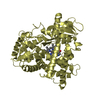
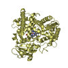
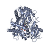
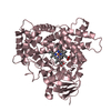
| Entry | Database: PDB / ID: 2fdv | ||||||
|---|---|---|---|---|---|---|---|
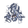
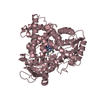
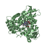
| Title | Microsomal P450 2A6 with the inhibitor N-Methyl(5-(pyridin-3-yl)furan-2-yl)methanamine bound | ||||||
 Components Components | Cytochrome P450 2A6 | ||||||
 Keywords Keywords | OXIDOREDUCTASE / CYP2A6 / P450 2A6 / P450 / MONOOXYGENASE / DRUG METABOLIZING ENZYME / COUMARIN 7-HYDROXYLASE / NICOTINE OXIDASE | ||||||
| Function / homology |  Function and homology information Function and homology informationcoumarin catabolic process / coumarin 7-hydroxylase activity / coumarin metabolic process / Oxidoreductases; Acting on paired donors, with incorporation or reduction of molecular oxygen; With reduced flavin or flavoprotein as one donor, and incorporation of one atom of oxygen into the other donor / CYP2E1 reactions / arachidonate epoxygenase activity / epoxygenase P450 pathway / Xenobiotics / oxidoreductase activity, acting on paired donors, with incorporation or reduction of molecular oxygen, reduced flavin or flavoprotein as one donor, and incorporation of one atom of oxygen / steroid metabolic process ...coumarin catabolic process / coumarin 7-hydroxylase activity / coumarin metabolic process / Oxidoreductases; Acting on paired donors, with incorporation or reduction of molecular oxygen; With reduced flavin or flavoprotein as one donor, and incorporation of one atom of oxygen into the other donor / CYP2E1 reactions / arachidonate epoxygenase activity / epoxygenase P450 pathway / Xenobiotics / oxidoreductase activity, acting on paired donors, with incorporation or reduction of molecular oxygen, reduced flavin or flavoprotein as one donor, and incorporation of one atom of oxygen / steroid metabolic process / xenobiotic catabolic process / cytoplasmic microtubule / xenobiotic metabolic process / iron ion binding / heme binding / endoplasmic reticulum membrane / enzyme binding / cytoplasm Similarity search - Function | ||||||
| Biological species |  Homo sapiens (human) Homo sapiens (human) | ||||||
| Method |  X-RAY DIFFRACTION / SYNCHROTRON / MOLECULAR REPLACEMENT / Resolution: 1.65 Å X-RAY DIFFRACTION / SYNCHROTRON / MOLECULAR REPLACEMENT / Resolution: 1.65 Å | ||||||
 Authors Authors | Yano, J.K. / Stout, C.D. / Johnson, E.F. | ||||||
 Citation Citation | Journal: J.Med.Chem. / Year: 2006 Title: Synthetic Inhibitors of Cytochrome P-450 2A6: Inhibitory Activity, Difference Spectra, Mechanism of Inhibition, and Protein Cocrystallization. Authors: Yano, J.K. / Denton, T.T. / Cerny, M.A. / Zhang, X. / Johnson, E.F. / Cashman, J.R. | ||||||
| History |
| ||||||
| Remark 999 | SEQUENCE RESIDUES 1-28 WERE REPLACED WITH THE SEQUENCE MAKKTS |
- Structure visualization
| Structure viewer | Molecule: MolmilJmol/JSmol |
|---|
- Downloads & links
-Download
| PDBx/mmCIF format | 2fdv.cif.gz | 414.8 KB | Display | PDBx/mmCIF format |
|---|---|---|---|---|
| PDB format | pdb2fdv.ent.gz | 335.9 KB | Display | PDB format |
| PDBx/mmJSON format | 2fdv.json.gz | Tree view | PDBx/mmJSON format | |
| Others |  Other downloads Other downloads |
-Validation report
| Arichive directory | https://data.pdbj.org/pub/pdb/validation_reports/fd/2fdvftp://data.pdbj.org/pub/pdb/validation_reports/fd/2fdv | HTTPS FTP |
|---|
-Related structure data
| Related structure data |  2fduC  2fdwC  2fdyC  1z10S S: Starting model for refinement C: citing same article ( |
|---|---|
| Similar structure data |


-Links
 PDBj
PDBj

- Assembly
| Deposited unit | 
| ||||||||
|---|---|---|---|---|---|---|---|---|---|
| 1 | 
| ||||||||
| 2 | 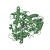
| ||||||||
| 3 | 
| ||||||||
| 4 | 
| ||||||||
| Unit cell |
|
-Components
-Protein , 1 types, 4 molecules ABCD
| #1: Protein | Mass: 54671.637 Da / Num. of mol.: 4 Source method: isolated from a genetically manipulated source Source: (gene. exp.) Homo sapiens (human) / Gene: CYP2A6 / Plasmid: PCWORI / Production host:  |
|---|
-Non-polymers , 5 types, 1457 molecules 


| #2: Chemical | ChemComp-SO4 /  Mass: 96.063 Da / Num. of mol.: 5 / Source method: obtained synthetically / Formula: SO4 Mass: 96.063 Da / Num. of mol.: 5 / Source method: obtained synthetically / Formula: SO4#3: Chemical | ChemComp-HEM /  Mass: 616.487 Da / Num. of mol.: 4 / Source method: obtained synthetically / Formula: C34H32FeN4O4 Mass: 616.487 Da / Num. of mol.: 4 / Source method: obtained synthetically / Formula: C34H32FeN4O4#4: Chemical | ChemComp-D2G /  Mass: 188.226 Da / Num. of mol.: 4 / Source method: obtained synthetically / Formula: C11H12N2O Mass: 188.226 Da / Num. of mol.: 4 / Source method: obtained synthetically / Formula: C11H12N2O#5: Chemical | ChemComp-EDO /  Mass: 62.068 Da / Num. of mol.: 7 / Source method: obtained synthetically / Formula: C2H6O2 Mass: 62.068 Da / Num. of mol.: 7 / Source method: obtained synthetically / Formula: C2H6O2#6: Water | ChemComp-HOH / | Mass: 18.015 Da / Num. of mol.: 1437 / Source method: isolated from a natural source / Formula: H2O |
|---|
-Experimental details
-Experiment
| Experiment | Method: X-RAY DIFFRACTION / Number of used crystals: 1 |
|---|
- Sample preparation
| Crystal | Density Matthews: 2.61 Å3/Da / Density % sol: 52.91 % |
|---|---|
| Crystal grow | Temperature: 298 K / Method: vapor diffusion, sitting drop / pH: 8.5 Details: Polyethylene Glycol2000, Monomethyl Ether, TRIS, AMMONIUM SULFATE, ANAPOE-X-405 , pH 8.5, VAPOR DIFFUSION, SITTING DROP, temperature 298K |
-Data collection
| Diffraction | Mean temperature: 100 K |
|---|---|
| Diffraction source | Source: SYNCHROTRON / Site: SSRL  / Beamline: BL9-1 / Wavelength: 0.98 Å / Beamline: BL9-1 / Wavelength: 0.98 Å |
| Detector | Type: ADSC QUANTUM 315 / Detector: CCD / Date: Mar 5, 2005 Details: Flat mirror (vertical focusing); single crystal Si(311) bent monochromator (horizontal focusing) |
| Radiation | Monochromator: single crystal Si(311) bent monochromator / Protocol: SINGLE WAVELENGTH / Monochromatic (M) / Laue (L): M / Scattering type: x-ray |
| Radiation wavelength | Wavelength: 0.98 Å / Relative weight: 1 |
| Reflection | Resolution: 1.6→33.8 Å / Num. all: 294239 / Num. obs: 278644 / % possible obs: 94.7 % / Observed criterion σ(F): 0 / Observed criterion σ(I): 0 / Redundancy: 3.5 % / Rsym value: 0.045 |
| Reflection shell | Resolution: 1.6→1.66 Å / Redundancy: 2.3 % / Mean I/σ(I) obs: 1.5 / Rsym value: 0.662 / % possible all: 70.1 |
- Processing
| Software |
| |||||||||||||||||||||||||
|---|---|---|---|---|---|---|---|---|---|---|---|---|---|---|---|---|---|---|---|---|---|---|---|---|---|---|
| Refinement | Method to determine structure: MOLECULAR REPLACEMENT Starting model: PDB ENTRY 1Z10 Resolution: 1.65→33.8 Å / Cross valid method: THROUGHOUT / σ(F): 0 / Stereochemistry target values: Engh & Huber
| |||||||||||||||||||||||||
| Refine analyze |
| |||||||||||||||||||||||||
| Refinement step | Cycle: LAST / Resolution: 1.65→33.8 Å
| |||||||||||||||||||||||||
| Refine LS restraints |
|
