 Movie
Movie Controller

[English] 日本語
 Yorodumi
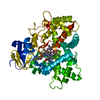
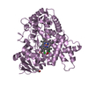
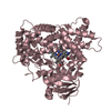
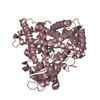
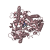
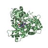
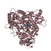
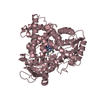
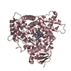
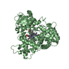
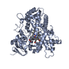
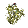
- PDB-2p85: Structure of Human Lung Cytochrome P450 2A13 with indole bound in... -
+ Open data

- Basic information
| Entry | Database: PDB / ID: 2p85 | ||||||
|---|---|---|---|---|---|---|---|
| Title | Structure of Human Lung Cytochrome P450 2A13 with indole bound in two alternate conformations | ||||||
 Components Components | Cytochrome P450 2A13 | ||||||
 Keywords Keywords | OXIDOREDUCTASE / CYP2A13 / P450 2A13 / P450 / monooxygenase / nicotine oxidase / coumarine 7-hydroxylase / NNK oxidase / 4-(methylnitrosamino)-1-(3-pyridyl)-1-butanone oxidase / heme | ||||||
| Function / homology |  Function and homology information Function and homology informationcoumarin 7-hydroxylase activity / Fatty acids / coumarin metabolic process / CYP2E1 reactions / arachidonate epoxygenase activity / epoxygenase P450 pathway / aflatoxin metabolic process / Aflatoxin activation and detoxification / Xenobiotics / oxidoreductase activity, acting on paired donors, with incorporation or reduction of molecular oxygen, reduced flavin or flavoprotein as one donor, and incorporation of one atom of oxygen ...coumarin 7-hydroxylase activity / Fatty acids / coumarin metabolic process / CYP2E1 reactions / arachidonate epoxygenase activity / epoxygenase P450 pathway / aflatoxin metabolic process / Aflatoxin activation and detoxification / Xenobiotics / oxidoreductase activity, acting on paired donors, with incorporation or reduction of molecular oxygen, reduced flavin or flavoprotein as one donor, and incorporation of one atom of oxygen / unspecific monooxygenase / xenobiotic metabolic process / monooxygenase activity / iron ion binding / heme binding / endoplasmic reticulum membrane / cytoplasm Similarity search - Function | ||||||
| Biological species |  Homo sapiens (human) Homo sapiens (human) | ||||||
| Method |  X-RAY DIFFRACTION / SYNCHROTRON / MOLECULAR REPLACEMENT / Resolution: 2.35 Å X-RAY DIFFRACTION / SYNCHROTRON / MOLECULAR REPLACEMENT / Resolution: 2.35 Å | ||||||
 Authors Authors | Scott, E.E. / Stout, C.D. | ||||||
 Citation Citation | Journal: J.Biol.Chem. / Year: 2007 Title: Structure of the human lung cytochrome P450 2A13. Authors: Smith, B.D. / Sanders, J.L. / Porubsky, P.R. / Lushington, G.H. / Stout, C.D. / Scott, E.E. | ||||||
| History |
|
- Structure visualization
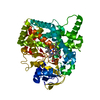
| Structure viewer | Molecule: MolmilJmol/JSmol |
|---|
- Downloads & links
-Download
| PDBx/mmCIF format | 2p85.cif.gz | 575 KB | Display | PDBx/mmCIF format |
|---|---|---|---|---|
| PDB format | pdb2p85.ent.gz | 475.2 KB | Display | PDB format |
| PDBx/mmJSON format | 2p85.json.gz | Tree view | PDBx/mmJSON format | |
| Others |  Other downloads Other downloads |
-Validation report
| Arichive directory | https://data.pdbj.org/pub/pdb/validation_reports/p8/2p85ftp://data.pdbj.org/pub/pdb/validation_reports/p8/2p85 | HTTPS FTP |
|---|
-Related structure data
| Related structure data | 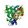 1suoS S: Starting model for refinement |
|---|---|
| Similar structure data |

-Links
 PDBj
PDBj


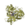
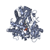
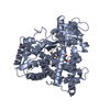
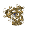
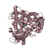
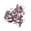
- Assembly
| Deposited unit | 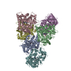
| ||||||||||||
|---|---|---|---|---|---|---|---|---|---|---|---|---|---|
| 1 | 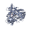
| ||||||||||||
| 2 | 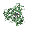
| ||||||||||||
| 3 | 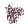
| ||||||||||||
| 4 | 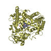
| ||||||||||||
| 5 | 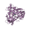
| ||||||||||||
| 6 | 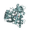
| ||||||||||||
| Unit cell |
| ||||||||||||
| Components on special symmetry positions |
|
-Components
| #1: Protein | Mass: 54804.758 Da / Num. of mol.: 6 / Fragment: catalytic domain (Residues 29-494) / Mutation: V23M, W24A, R25K, Q26K, R27T, K28S, R30K Source method: isolated from a genetically manipulated source Source: (gene. exp.) Homo sapiens (human) / Gene: CYP2A13 / Plasmid: pKK2A13dH / Production host:  #2: Chemical | ChemComp-HEM /   Mass: 616.487 Da / Num. of mol.: 6 / Source method: obtained synthetically / Formula: C34H32FeN4O4 Mass: 616.487 Da / Num. of mol.: 6 / Source method: obtained synthetically / Formula: C34H32FeN4O4#3: Chemical | ChemComp-IND / 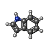  Mass: 117.148 Da / Num. of mol.: 12 / Source method: obtained synthetically / Formula: C8H7N Mass: 117.148 Da / Num. of mol.: 12 / Source method: obtained synthetically / Formula: C8H7N#4: Water | ChemComp-HOH / |  Mass: 18.015 Da / Num. of mol.: 597 / Source method: isolated from a natural source / Formula: H2O Mass: 18.015 Da / Num. of mol.: 597 / Source method: isolated from a natural source / Formula: H2O |
|---|
-Experimental details
-Experiment
| Experiment | Method: X-RAY DIFFRACTION / Number of used crystals: 1 |
|---|
- Sample preparation
| Crystal | Density Matthews: 2.71 Å3/Da / Density % sol: 54.58 % |
|---|---|
| Crystal grow | Temperature: 293 K / Method: vapor diffusion, hanging drop Details: Ammonium Sulfate, Hepes, PEG 2000 monomethyl ether , VAPOR DIFFUSION, HANGING DROP, temperature 293K |
-Data collection
| Diffraction | Mean temperature: 100 K |
|---|---|
| Diffraction source | Source: SYNCHROTRON / Site: SSRL  / Beamline: BL11-1 / Wavelength: 0.98 Å / Beamline: BL11-1 / Wavelength: 0.98 Å |
| Detector | Type: ADSC QUANTUM 315 / Detector: CCD / Date: Apr 22, 2005 |
| Radiation | Monochromator: SI(111) / Protocol: SINGLE WAVELENGTH / Monochromatic (M) / Laue (L): M / Scattering type: x-ray |
| Radiation wavelength | Wavelength: 0.98 Å / Relative weight: 1 |
| Reflection | Highest resolution: 2.35 Å / Num. obs: 142896 / Redundancy: 4.7 % / Biso Wilson estimate: 36.2 Å2 / Net I/σ(I): 14.5 |
| Reflection shell | Highest resolution: 2.35 Å / Redundancy: 3.7 % / Mean I/σ(I) obs: 2.9 / Rsym value: 0.431 / % possible all: 97.3 |
- Processing
| Software |
| ||||||||||||||||||||||||||||||||||||||||||||||||||||||||||||||||||||||||||||||||
|---|---|---|---|---|---|---|---|---|---|---|---|---|---|---|---|---|---|---|---|---|---|---|---|---|---|---|---|---|---|---|---|---|---|---|---|---|---|---|---|---|---|---|---|---|---|---|---|---|---|---|---|---|---|---|---|---|---|---|---|---|---|---|---|---|---|---|---|---|---|---|---|---|---|---|---|---|---|---|---|---|---|
| Refinement | Method to determine structure: MOLECULAR REPLACEMENT Starting model: 1SUO Resolution: 2.35→29.34 Å / Rfactor Rfree error: 0.002 / Data cutoff high absF: 1849573.99 / Data cutoff low absF: 0 / Isotropic thermal model: RESTRAINED / Cross valid method: THROUGHOUT / σ(F): 0 / Stereochemistry target values: Engh & Huber
| ||||||||||||||||||||||||||||||||||||||||||||||||||||||||||||||||||||||||||||||||
| Solvent computation | Solvent model: FLAT MODEL / Bsol: 46.6015 Å2 / ksol: 0.353435 e/Å3 | ||||||||||||||||||||||||||||||||||||||||||||||||||||||||||||||||||||||||||||||||
| Displacement parameters | Biso mean: 45.5 Å2
| ||||||||||||||||||||||||||||||||||||||||||||||||||||||||||||||||||||||||||||||||
| Refine analyze |
| ||||||||||||||||||||||||||||||||||||||||||||||||||||||||||||||||||||||||||||||||
| Refinement step | Cycle: LAST / Resolution: 2.35→29.34 Å
| ||||||||||||||||||||||||||||||||||||||||||||||||||||||||||||||||||||||||||||||||
| Refine LS restraints |
| ||||||||||||||||||||||||||||||||||||||||||||||||||||||||||||||||||||||||||||||||
| LS refinement shell | Resolution: 2.35→2.5 Å / Rfactor Rfree error: 0.007 / Total num. of bins used: 6
| ||||||||||||||||||||||||||||||||||||||||||||||||||||||||||||||||||||||||||||||||
| Xplor file |
|
