 Movie
Movie Controller

[English] 日本語
 Yorodumi
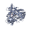
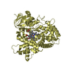
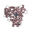
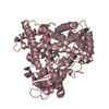
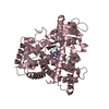
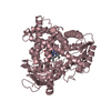
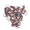
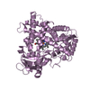
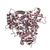
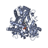
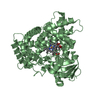
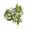
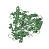
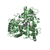
- PDB-4eji: Human Cytochrome P450 2A13 in complex with two molecules of 4-(me... -
+ Open data

- Basic information
| Entry | Database: PDB / ID: 4eji | ||||||
|---|---|---|---|---|---|---|---|
| Title | Human Cytochrome P450 2A13 in complex with two molecules of 4-(methylnitrosamino)-1-(3-puridyl)-1-butanone | ||||||
 Components Components | Cytochrome P450 2A13 | ||||||
 Keywords Keywords | OXIDOREDUCTASE / CYP2A13 / cytochrome P450 2A13 / P450 2A13 / heme protein / monooxygenase / drug metabolism / xenobiotic metabolism / endoplasmic reticulum / membrane | ||||||
| Function / homology |  Function and homology information Function and homology informationcoumarin 7-hydroxylase activity / Fatty acids / coumarin metabolic process / CYP2E1 reactions / arachidonate epoxygenase activity / epoxygenase P450 pathway / aflatoxin metabolic process / Aflatoxin activation and detoxification / Xenobiotics / oxidoreductase activity, acting on paired donors, with incorporation or reduction of molecular oxygen, reduced flavin or flavoprotein as one donor, and incorporation of one atom of oxygen ...coumarin 7-hydroxylase activity / Fatty acids / coumarin metabolic process / CYP2E1 reactions / arachidonate epoxygenase activity / epoxygenase P450 pathway / aflatoxin metabolic process / Aflatoxin activation and detoxification / Xenobiotics / oxidoreductase activity, acting on paired donors, with incorporation or reduction of molecular oxygen, reduced flavin or flavoprotein as one donor, and incorporation of one atom of oxygen / unspecific monooxygenase / xenobiotic metabolic process / monooxygenase activity / iron ion binding / heme binding / endoplasmic reticulum membrane / cytoplasm Similarity search - Function | ||||||
| Biological species |  Homo sapiens (human) Homo sapiens (human) | ||||||
| Method |  X-RAY DIFFRACTION / SYNCHROTRON / MOLECULAR REPLACEMENT / Resolution: 2.1 Å X-RAY DIFFRACTION / SYNCHROTRON / MOLECULAR REPLACEMENT / Resolution: 2.1 Å | ||||||
 Authors Authors | DeVore, N.M. / Scott, E.E. | ||||||
 Citation Citation | Journal: J.Biol.Chem. / Year: 2012 Title: Nicotine and 4-(methylnitrosamino)-1-(3-pyridyl)-1-butanone binding and access channel in human cytochrome P450 2A6 and 2A13 enzymes. Authors: DeVore, N.M. / Scott, E.E. | ||||||
| History |
|
- Structure visualization
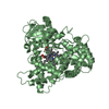
| Structure viewer | Molecule: MolmilJmol/JSmol |
|---|
- Downloads & links
-Download
| PDBx/mmCIF format | 4eji.cif.gz | 110.9 KB | Display | PDBx/mmCIF format |
|---|---|---|---|---|
| PDB format | pdb4eji.ent.gz | 84.1 KB | Display | PDB format |
| PDBx/mmJSON format | 4eji.json.gz | Tree view | PDBx/mmJSON format | |
| Others |  Other downloads Other downloads |
-Validation report
| Arichive directory | https://data.pdbj.org/pub/pdb/validation_reports/ej/4ejiftp://data.pdbj.org/pub/pdb/validation_reports/ej/4eji | HTTPS FTP |
|---|
-Related structure data
| Related structure data |  4ejgC  4ejhC 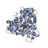 4ejjC  2p85S S: Starting model for refinement C: citing same article ( |
|---|---|
| Similar structure data |


-Links
 PDBj
PDBj


- Assembly
| Deposited unit | 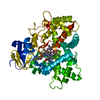
| ||||||||
|---|---|---|---|---|---|---|---|---|---|
| 1 |
| ||||||||
| Unit cell |
|
-Components
| #1: Protein | Mass: 54804.758 Da / Num. of mol.: 1 / Fragment: unp residues 31-494 Source method: isolated from a genetically manipulated source Source: (gene. exp.) Homo sapiens (human) / Gene: CYP2A13 / Plasmid: pKK2A13dH / Production host:  | ||
|---|---|---|---|
| #2: Chemical | ChemComp-HEM /   Mass: 616.487 Da / Num. of mol.: 1 / Source method: obtained synthetically / Formula: C34H32FeN4O4 Mass: 616.487 Da / Num. of mol.: 1 / Source method: obtained synthetically / Formula: C34H32FeN4O4 | ||
| #3: Chemical |   Mass: 207.229 Da / Num. of mol.: 2 / Source method: obtained synthetically / Formula: C10H13N3O2 Mass: 207.229 Da / Num. of mol.: 2 / Source method: obtained synthetically / Formula: C10H13N3O2#4: Water | ChemComp-HOH / |  Mass: 18.015 Da / Num. of mol.: 69 / Source method: isolated from a natural source / Formula: H2O Mass: 18.015 Da / Num. of mol.: 69 / Source method: isolated from a natural source / Formula: H2O |
-Experimental details
-Experiment
| Experiment | Method: X-RAY DIFFRACTION / Number of used crystals: 1 |
|---|
- Sample preparation
| Crystal | Density Matthews: 3.34 Å3/Da / Density % sol: 63.18 % |
|---|---|
| Crystal grow | Temperature: 298 K / pH: 7.8 Details: 4.0 M sodium formate, pH 7.8, VAPOR DIFFUSION, HANGING DROP, temperature 298K |
-Data collection
| Diffraction | Mean temperature: 100 K |
|---|---|
| Diffraction source | Source: SYNCHROTRON / Site: SSRL  / Beamline: BL9-2 / Wavelength: 0.98 / Beamline: BL9-2 / Wavelength: 0.98 |
| Detector | Type: MARMOSAIC 325 mm CCD / Detector: CCD / Date: Jan 27, 2012 / Details: RH COAT FLAT MIRROR, TOROIDAL FOCUSING MIRROR |
| Radiation | Monochromator: DOUBLE CRYSTAL MONOCHROMATOR / Protocol: SINGLE WAVELENGTH / Monochromatic (M) / Laue (L): M / Scattering type: x-ray |
| Radiation wavelength | Wavelength: 0.98 Å / Relative weight: 1 |
| Reflection | Resolution: 2.1→97.11 Å / Num. obs: 43580 / % possible obs: 100 % / Observed criterion σ(I): 0 / Redundancy: 14.7 % / Biso Wilson estimate: 41.1 Å2 / Rmerge(I) obs: 0.101 / Rsym value: 0.104 / Net I/σ(I): 18.3 |
| Reflection shell | Resolution: 2.1→2.15 Å / Redundancy: 14.8 % / Mean I/σ(I) obs: 2.1 / % possible all: 100 |
- Processing
| Software |
| |||||||||||||||||||||||||
|---|---|---|---|---|---|---|---|---|---|---|---|---|---|---|---|---|---|---|---|---|---|---|---|---|---|---|
| Refinement | Method to determine structure: MOLECULAR REPLACEMENT Starting model: PDB ENTRY 2P85 Resolution: 2.1→51.9 Å / Cor.coef. Fo:Fc: 0.958 / Cor.coef. Fo:Fc free: 0.947 / SU B: 4.629 / SU ML: 0.12 / Cross valid method: THROUGHOUT / σ(F): 0 / ESU R: 0.168 / ESU R Free: 0.155 Stereochemistry target values: MAXIMUM LIKELIHOOD WITH PHASES
| |||||||||||||||||||||||||
| Solvent computation | Ion probe radii: 0.8 Å / Shrinkage radii: 0.8 Å / VDW probe radii: 1.4 Å / Solvent model: MASK | |||||||||||||||||||||||||
| Displacement parameters | Biso mean: 40.98 Å2
| |||||||||||||||||||||||||
| Refinement step | Cycle: LAST / Resolution: 2.1→51.9 Å
| |||||||||||||||||||||||||
| LS refinement shell | Resolution: 2.1→2.154 Å / Total num. of bins used: 20
|
