- PDB-4i91: Crystal Structure of Cytochrome P450 2B6 (Y226H/K262R) in complex... -
+
Open data
ID or keywords:
Loading...
-
Basic information
Entry
Database: PDB / ID: 4i91
Title
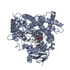
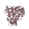
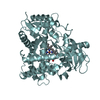
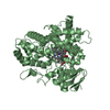
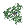
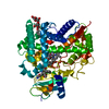
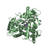
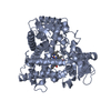
Crystal Structure of Cytochrome P450 2B6 (Y226H/K262R) in complex with alpha-Pinene.
Components
Cytochrome P450 2B6
Keywords
OXIDOREDUCTASE / membrane protein / CYP2B6 / P450 / Cytochrome P450 2B6 / Monooxygenase / Endoplasmic Reticulum / Heme / Iron / Membrane / Metal Binding / Microsome
Function / homology
Function and homology information
testosterone 16-alpha-hydroxylase activity / testosterone 16-beta-hydroxylase activity / Fatty acids / Oxidoreductases; Acting on paired donors, with incorporation or reduction of molecular oxygen; With NADH or NADPH as one donor, and incorporation of one atom of oxygen into the other donor / CYP2E1 reactions / arachidonate epoxygenase activity / ketone metabolic process / epoxygenase P450 pathway / anandamide 8,9 epoxidase activity / anandamide 11,12 epoxidase activity ...testosterone 16-alpha-hydroxylase activity / testosterone 16-beta-hydroxylase activity / Fatty acids / Oxidoreductases; Acting on paired donors, with incorporation or reduction of molecular oxygen; With NADH or NADPH as one donor, and incorporation of one atom of oxygen into the other donor / CYP2E1 reactions / arachidonate epoxygenase activity / ketone metabolic process / epoxygenase P450 pathway / anandamide 8,9 epoxidase activity / anandamide 11,12 epoxidase activity / anandamide 14,15 epoxidase activity / estrogen 2-hydroxylase activity / Xenobiotics / Phase I - Functionalization of compounds / oxidoreductase activity, acting on paired donors, with incorporation or reduction of molecular oxygen, reduced flavin or flavoprotein as one donor, and incorporation of one atom of oxygen / steroid metabolic process / xenobiotic catabolic process / xenobiotic metabolic process / monooxygenase activity / iron ion binding / heme binding / endoplasmic reticulum membrane / cytoplasm Similarity search - Function
Cytochrome P450, E-class, group I, CYP2B-like / : / Cytochrome P450, E-class, group I / Cytochrome p450 / Cytochrome P450 / Cytochrome P450, conserved site / Cytochrome P450 cysteine heme-iron ligand signature. / Cytochrome P450 / Cytochrome P450 superfamily / Cytochrome P450 ...Cytochrome P450, E-class, group I, CYP2B-like / : / Cytochrome P450, E-class, group I / Cytochrome p450 / Cytochrome P450 / Cytochrome P450, conserved site / Cytochrome P450 cysteine heme-iron ligand signature. / Cytochrome P450 / Cytochrome P450 superfamily / Cytochrome P450 / Orthogonal Bundle / Mainly Alpha Similarity search - Domain/homology
beta-D-fructofuranose / PROTOPORPHYRIN IX CONTAINING FE / (+)-alpha-Pinene / Cytochrome P450 2B6 Similarity search - Component
Mass: 54646.004 Da / Num. of mol.: 1 / Fragment: Cytochrome P450 2B6 / Mutation: Y226H, K262R Source method: isolated from a genetically manipulated source Source: (gene. exp.) Homo sapiens (human) / Gene: CYP2B6 / Plasmid: pKK / Production host: Escherichia coli (E. coli) / Strain (production host): JM109 References: UniProt: P20813, Oxidoreductases; Acting on paired donors, with incorporation or reduction of molecular oxygen; With NADH or NADPH as one donor, and incorporation of one atom of oxygen into the other donor
Mass: 18.015 Da / Num. of mol.: 369 / Source method: isolated from a natural source / Formula: H2O
-
Details
Sequence details
THE RESIDUES 3-21 (LSVLLFLALLTGLLLLLVQ) OF CORRESPONDING DATABASE REFERENCE SEQUENCE (UNP P20813) ...THE RESIDUES 3-21 (LSVLLFLALLTGLLLLLVQ) OF CORRESPONDING DATABASE REFERENCE SEQUENCE (UNP P20813) ARE DELETED IN THIS STRUCTURE.
-
Experimental details
-
Experiment
Experiment
Method: X-RAY DIFFRACTION / Number of used crystals: 1
-
Sample preparation
Crystal
Density Matthews: 3.17 Å3/Da / Density % sol: 61.15 %
Crystal grow
Temperature: 291 K / Method: vapor diffusion, sitting drop / pH: 7.5 Details: 0.1 M HEPES sodium pH 7.5 and 1.4 M sodium citrate tribasic dihydrate, VAPOR DIFFUSION, SITTING DROP, temperature 291K
Resolution: 2→20 Å / Cor.coef. Fo:Fc: 0.954 / Cor.coef. Fo:Fc free: 0.94 / SU B: 3.081 / SU ML: 0.087 / Cross valid method: THROUGHOUT / ESU R: 0.143 / ESU R Free: 0.131 / Stereochemistry target values: MAXIMUM LIKELIHOOD / Details: HYDROGENS HAVE BEEN ADDED IN THE RIDING POSITIONS
Rfactor
Num. reflection
% reflection
Selection details
Rfree
0.21001
2418
5.1 %
RANDOM
Rwork
0.18119
-
-
-
obs
0.18262
45252
99.66 %
-
all
-
45406
-
-
Solvent computation
Ion probe radii: 0.8 Å / Shrinkage radii: 0.8 Å / VDW probe radii: 1.4 Å / Solvent model: MASK
Displacement parameters
Biso mean: 26.129 Å2
Baniso -1
Baniso -2
Baniso -3
1-
0.31 Å2
0.15 Å2
-0 Å2
2-
-
0.31 Å2
-0 Å2
3-
-
-
-0.46 Å2
Refinement step
Cycle: LAST / Resolution: 2→20 Å
Protein
Nucleic acid
Ligand
Solvent
Total
Num. atoms
3757
0
123
369
4249
Refine LS restraints
Refine-ID
Type
Dev ideal
Dev ideal target
Number
X-RAY DIFFRACTION
r_bond_refined_d
0.01
0.022
3992
X-RAY DIFFRACTION
r_bond_other_d
X-RAY DIFFRACTION
r_angle_refined_deg
1.303
2.023
5411
X-RAY DIFFRACTION
r_angle_other_deg
X-RAY DIFFRACTION
r_dihedral_angle_1_deg
5.018
5
464
X-RAY DIFFRACTION
r_dihedral_angle_2_deg
34.501
22.826
184
X-RAY DIFFRACTION
r_dihedral_angle_3_deg
12.609
15
661
X-RAY DIFFRACTION
r_dihedral_angle_4_deg
16.865
15
30
X-RAY DIFFRACTION
r_chiral_restr
0.27
0.2
582
X-RAY DIFFRACTION
r_gen_planes_refined
0.005
0.021
3008
X-RAY DIFFRACTION
r_gen_planes_other
X-RAY DIFFRACTION
r_nbd_refined
X-RAY DIFFRACTION
r_nbd_other
X-RAY DIFFRACTION
r_nbtor_refined
X-RAY DIFFRACTION
r_nbtor_other
X-RAY DIFFRACTION
r_xyhbond_nbd_refined
X-RAY DIFFRACTION
r_xyhbond_nbd_other
X-RAY DIFFRACTION
r_metal_ion_refined
X-RAY DIFFRACTION
r_metal_ion_other
X-RAY DIFFRACTION
r_symmetry_vdw_refined
X-RAY DIFFRACTION
r_symmetry_vdw_other
X-RAY DIFFRACTION
r_symmetry_hbond_refined
X-RAY DIFFRACTION
r_symmetry_hbond_other
X-RAY DIFFRACTION
r_symmetry_metal_ion_refined
X-RAY DIFFRACTION
r_symmetry_metal_ion_other
X-RAY DIFFRACTION
r_mcbond_it
0.542
1.5
2323
X-RAY DIFFRACTION
r_mcbond_other
X-RAY DIFFRACTION
r_mcangle_it
1.062
2
3763
X-RAY DIFFRACTION
r_scbond_it
1.591
3
1669
X-RAY DIFFRACTION
r_scangle_it
2.703
4.5
1646
X-RAY DIFFRACTION
r_rigid_bond_restr
X-RAY DIFFRACTION
r_sphericity_free
X-RAY DIFFRACTION
r_sphericity_bonded
LS refinement shell
Resolution: 2→2.051 Å / Total num. of bins used: 20
Rfactor
Num. reflection
% reflection
Rfree
0.253
189
-
Rwork
0.226
3194
-
obs
-
-
98.54 %
+
About Yorodumi
-
News
-
Feb 9, 2022. New format data for meta-information of EMDB entries
New format data for meta-information of EMDB entries
Version 3 of the EMDB header file is now the official format.
The previous official version 1.9 will be removed from the archive.
In the structure databanks used in Yorodumi, some data are registered as the other names, "COVID-19 virus" and "2019-nCoV". Here are the details of the virus and the list of structure data.
Jan 31, 2019. EMDB accession codes are about to change! (news from PDBe EMDB page)
EMDB accession codes are about to change! (news from PDBe EMDB page)
The allocation of 4 digits for EMDB accession codes will soon come to an end. Whilst these codes will remain in use, new EMDB accession codes will include an additional digit and will expand incrementally as the available range of codes is exhausted. The current 4-digit format prefixed with “EMD-” (i.e. EMD-XXXX) will advance to a 5-digit format (i.e. EMD-XXXXX), and so on. It is currently estimated that the 4-digit codes will be depleted around Spring 2019, at which point the 5-digit format will come into force.
The EM Navigator/Yorodumi systems omit the EMD- prefix.
Related info.:Q: What is EMD? / ID/Accession-code notation in Yorodumi/EM Navigator
Yorodumi is a browser for structure data from EMDB, PDB, SASBDB, etc.
This page is also the successor to EM Navigator detail page, and also detail information page/front-end page for Omokage search.
The word "yorodu" (or yorozu) is an old Japanese word meaning "ten thousand". "mi" (miru) is to see.
Related info.:EMDB / PDB / SASBDB / Comparison of 3 databanks / Yorodumi Search / Aug 31, 2016. New EM Navigator & Yorodumi / Yorodumi Papers / Jmol/JSmol / Function and homology information / Changes in new EM Navigator and Yorodumi
 Movie
Movie Controller

 Yorodumi
 Open data
 Basic information
 Components
 Keywords
 Function and homology information
 Homo sapiens (human)
 X-RAY DIFFRACTION /
  Authors
 Citation
 Structure visualization
 Downloads & links
 Other downloads


 PDBj
PDBj


 Assembly
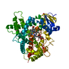

 Type: D-saccharide, beta linking / Mass: 180.156 Da / Num. of mol.: 1


 Mass: 616.487 Da / Num. of mol.: 1 / Source method: obtained synthetically / Formula: C34H32FeN4O4
 Mass: 494.573 Da / Num. of mol.: 3 / Source method: obtained synthetically / Formula: C23H42O11 / Comment: detergent*YM
 Mass: 136.234 Da / Num. of mol.: 1 / Source method: obtained synthetically / Formula: C10H16
 Sample preparation
 / Beamline: BL7-1 / Wavelength: 0.97 Å
 Processing