testosterone 16-alpha-hydroxylase activity / testosterone 16-beta-hydroxylase activity / Fatty acids / Oxidoreductases; Acting on paired donors, with incorporation or reduction of molecular oxygen; With NADH or NADPH as one donor, and incorporation of one atom of oxygen into the other donor / CYP2E1 reactions / arachidonate epoxygenase activity / ketone metabolic process / epoxygenase P450 pathway / anandamide 8,9 epoxidase activity / anandamide 11,12 epoxidase activity ...testosterone 16-alpha-hydroxylase activity / testosterone 16-beta-hydroxylase activity / Fatty acids / Oxidoreductases; Acting on paired donors, with incorporation or reduction of molecular oxygen; With NADH or NADPH as one donor, and incorporation of one atom of oxygen into the other donor / CYP2E1 reactions / arachidonate epoxygenase activity / ketone metabolic process / epoxygenase P450 pathway / anandamide 8,9 epoxidase activity / anandamide 11,12 epoxidase activity / anandamide 14,15 epoxidase activity / estrogen 2-hydroxylase activity / Xenobiotics / Phase I - Functionalization of compounds / oxidoreductase activity, acting on paired donors, with incorporation or reduction of molecular oxygen, reduced flavin or flavoprotein as one donor, and incorporation of one atom of oxygen / steroid metabolic process / xenobiotic catabolic process / xenobiotic metabolic process / monooxygenase activity / iron ion binding / heme binding / endoplasmic reticulum membrane / cytoplasm Similarity search - Function
Cytochrome P450, E-class, group I, CYP2B-like / : / Cytochrome P450, E-class, group I / Cytochrome p450 / Cytochrome P450 / Cytochrome P450, conserved site / Cytochrome P450 cysteine heme-iron ligand signature. / Cytochrome P450 / Cytochrome P450 superfamily / Cytochrome P450 ...Cytochrome P450, E-class, group I, CYP2B-like / : / Cytochrome P450, E-class, group I / Cytochrome p450 / Cytochrome P450 / Cytochrome P450, conserved site / Cytochrome P450 cysteine heme-iron ligand signature. / Cytochrome P450 / Cytochrome P450 superfamily / Cytochrome P450 / Orthogonal Bundle / Mainly Alpha Similarity search - Domain/homology
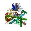
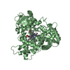
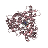
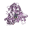
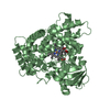
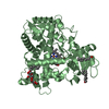
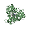
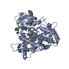
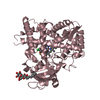
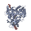
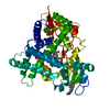
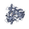
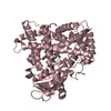
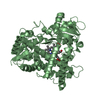
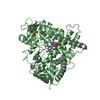
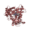
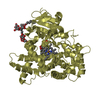
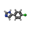
4-(4-CHLOROPHENYL)IMIDAZOLE / PROTOPORPHYRIN IX CONTAINING FE / THIOCYANATE ION / Cytochrome P450 2B6 Similarity search - Component
Type: MARMOSAIC 325 mm CCD / Detector: CCD / Date: Jun 19, 2009
Radiation
Monochromator: SI(111) / Protocol: SINGLE WAVELENGTH / Monochromatic (M) / Laue (L): M / Scattering type: x-ray
Radiation wavelength
Wavelength: 0.98 Å / Relative weight: 1
Reflection
Redundancy: 6.9 % / Av σ(I) over netI: 4.6 / Number: 223616 / Rmerge(I) obs: 0.131 / Rsym value: 0.131 / D res high: 1.998 Å / D res low: 98.451 Å / Num. obs: 32536 / % possible obs: 98.3
Resolution: 2→2.11 Å / Redundancy: 4.1 % / Rmerge(I) obs: 0.487 / Mean I/σ(I) obs: 1.4 / Rsym value: 0.487 / % possible all: 88.1
-
Phasing
Phasing
Method: molecular replacement
Phasing MR
Model details: Phaser MODE: MR_AUTO
Highest resolution
Lowest resolution
Rotation
2.5 Å
49.23 Å
Translation
2.5 Å
49.23 Å
-
Processing
Software
Name
Version
Classification
NB
SCALA
3.2.25
datascaling
PHASER
1.3.3
phasing
REFMAC
refinement
PDB_EXTRACT
3.005
dataextraction
MOSFLM
datareduction
Refinement
Method to determine structure: MOLECULAR REPLACEMENT Starting model: ENSEMBLE OF PDB ID 2Q6N (A CHAIN), 1SUO, 2BDM, 3G5N (A CHAIN) EACH WITH RESIDUES 101-120, 205-232 OMITTED
In the structure databanks used in Yorodumi, some data are registered as the other names, "COVID-19 virus" and "2019-nCoV". Here are the details of the virus and the list of structure data.
Jan 31, 2019. EMDB accession codes are about to change! (news from PDBe EMDB page)
EMDB accession codes are about to change! (news from PDBe EMDB page)
The allocation of 4 digits for EMDB accession codes will soon come to an end. Whilst these codes will remain in use, new EMDB accession codes will include an additional digit and will expand incrementally as the available range of codes is exhausted. The current 4-digit format prefixed with “EMD-” (i.e. EMD-XXXX) will advance to a 5-digit format (i.e. EMD-XXXXX), and so on. It is currently estimated that the 4-digit codes will be depleted around Spring 2019, at which point the 5-digit format will come into force.
The EM Navigator/Yorodumi systems omit the EMD- prefix.
Related info.:Q: What is EMD? / ID/Accession-code notation in Yorodumi/EM Navigator
Yorodumi is a browser for structure data from EMDB, PDB, SASBDB, etc.
This page is also the successor to EM Navigator detail page, and also detail information page/front-end page for Omokage search.
The word "yorodu" (or yorozu) is an old Japanese word meaning "ten thousand". "mi" (miru) is to see.
Related info.:EMDB / PDB / SASBDB / Comparison of 3 databanks / Yorodumi Search / Aug 31, 2016. New EM Navigator & Yorodumi / Yorodumi Papers / Jmol/JSmol / Function and homology information / Changes in new EM Navigator and Yorodumi
 Movie
Movie Controller

 Yorodumi
 Open data
 Basic information
 Components
 Keywords
 Function and homology information
 Homo sapiens (human)
 X-RAY DIFFRACTION /
  Authors
 Citation
 Structure visualization
 Downloads & links
 Other downloads
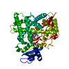


 PDBj
PDBj


 Assembly
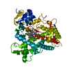


 Mass: 616.487 Da / Num. of mol.: 1 / Source method: obtained synthetically / Formula: C34H32FeN4O4
 Mass: 178.618 Da / Num. of mol.: 1 / Source method: obtained synthetically / Formula: C9H7ClN2
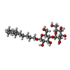
 Mass: 494.573 Da / Num. of mol.: 2 / Source method: obtained synthetically / Formula: C23H42O11 / Comment: detergent*YM
 Mass: 58.082 Da / Num. of mol.: 4 / Source method: obtained synthetically / Formula: CNS
 Sample preparation
 / Beamline: BL11-1 / Wavelength: 0.98
  Processing