 Movie
Movie Controller


+ Open data

- Basic information
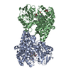
| Entry | Database: PDB / ID: 1k9x | ||||||
|---|---|---|---|---|---|---|---|
| Title | Structure of Pyrococcus furiosus carboxypeptidase Apo-Yb | ||||||
 Components Components | M32 carboxypeptidase | ||||||
 Keywords Keywords | carboxypeptidase / HEXXH motif / M32 family / metallopeptidase | ||||||
| Function / homology |  Function and homology information Function and homology informationcarboxypeptidase Taq / cobalt ion binding / metallocarboxypeptidase activity / proteolysis Similarity search - Function | ||||||
| Biological species |   Pyrococcus furiosus (archaea) Pyrococcus furiosus (archaea) | ||||||
| Method |  X-RAY DIFFRACTION / SYNCHROTRON / MAD / Resolution: 2.3 Å X-RAY DIFFRACTION / SYNCHROTRON / MAD / Resolution: 2.3 Å | ||||||
 Authors Authors | Arndt, J.W. / Hao, B. / Ramakrishnan, V. / Cheng, T. / Chan, S.I. / Chan, M.K. | ||||||
 Citation Citation | Journal: Structure / Year: 2002 Title: Crystal Structure of a Novel Carboxypeptidase from the Hyperthermophilic Archaeon Pyrococcus furiosus Authors: Arndt, J.W. / Hao, B. / Ramakrishnan, V. / Cheng, T. / Chan, S.I. / Chan, M.K. | ||||||
| History |
| ||||||
| Remark 999 | SEQUENCE An appropriate sequence database reference was not available at the time of processing. |
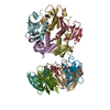
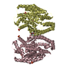
- Structure visualization
| Structure viewer | Molecule: MolmilJmol/JSmol |
|---|
- Downloads & links
-Download
| PDBx/mmCIF format | 1k9x.cif.gz | 412.5 KB | Display | PDBx/mmCIF format |
|---|---|---|---|---|
| PDB format | pdb1k9x.ent.gz | 341.6 KB | Display | PDB format |
| PDBx/mmJSON format | 1k9x.json.gz | Tree view | PDBx/mmJSON format | |
| Others |  Other downloads Other downloads |
-Validation report
| Arichive directory | https://data.pdbj.org/pub/pdb/validation_reports/k9/1k9xftp://data.pdbj.org/pub/pdb/validation_reports/k9/1k9x | HTTPS FTP |
|---|
-Related structure data


-Links
 PDBj
PDBj- Assembly
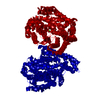
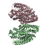
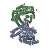
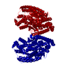
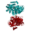
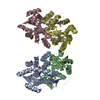
| Deposited unit | 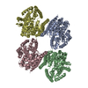
| ||||||||
|---|---|---|---|---|---|---|---|---|---|
| 1 | 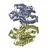
| ||||||||
| 2 | 
| ||||||||
| Unit cell |
|
-Components
| #1: Protein | Mass: 59127.664 Da / Num. of mol.: 4 / Source method: isolated from a natural source / Source: (natural) Pyrococcus furiosus (archaea) / References: UniProt: Q8U3L0#2: Water | ChemComp-HOH / |  Mass: 18.015 Da / Num. of mol.: 609 / Source method: isolated from a natural source / Formula: H2O Mass: 18.015 Da / Num. of mol.: 609 / Source method: isolated from a natural source / Formula: H2O |
|---|
-Experimental details
-Experiment
| Experiment | Method: X-RAY DIFFRACTION / Number of used crystals: 1 |
|---|
- Sample preparation
| Crystal | Density Matthews: 2.45 Å3/Da / Density % sol: 49.38 % | |||||||||||||||||||||||||||||||||||||||||||||||||
|---|---|---|---|---|---|---|---|---|---|---|---|---|---|---|---|---|---|---|---|---|---|---|---|---|---|---|---|---|---|---|---|---|---|---|---|---|---|---|---|---|---|---|---|---|---|---|---|---|---|---|
| Crystal grow | Temperature: 277 K / Method: vapor diffusion, hanging drop / pH: 8.5 Details: PEG 4000, TRIS, magnesium chloride, pH 8.5, VAPOR DIFFUSION, HANGING DROP, temperature 277K | |||||||||||||||||||||||||||||||||||||||||||||||||
| Crystal grow | *PLUS Temperature: 4 ℃ / pH: 8 | |||||||||||||||||||||||||||||||||||||||||||||||||
| Components of the solutions | *PLUS
|
-Data collection
| Diffraction | Mean temperature: 100 K | |||||||||
|---|---|---|---|---|---|---|---|---|---|---|
| Diffraction source | Source: SYNCHROTRON / Site: SSRL  / Beamline: BL9-2 / Wavelength: 1.0000, 1.3860 / Beamline: BL9-2 / Wavelength: 1.0000, 1.3860 | |||||||||
| Detector | Type: ADSC QUANTUM 4 / Detector: CCD / Date: Jan 1, 2000 | |||||||||
| Radiation | Monochromator: double crystal / Protocol: MAD / Monochromatic (M) / Laue (L): M / Scattering type: x-ray | |||||||||
| Radiation wavelength |
| |||||||||
| Reflection | Resolution: 2.3→50 Å / Num. all: 98094 / Num. obs: 98094 / % possible obs: 93.8 % / Observed criterion σ(F): 2 / Observed criterion σ(I): 2 / Biso Wilson estimate: 20.2 Å2 / Rmerge(I) obs: 0.036 / Net I/σ(I): 26.2 | |||||||||
| Reflection shell | Resolution: 2.3→2.34 Å / Rmerge(I) obs: 0.087 / % possible all: 80.5 | |||||||||
| Reflection | *PLUS Lowest resolution: 50 Å / Num. measured all: 353793 / Rmerge(I) obs: 0.036 | |||||||||
| Reflection shell | *PLUS % possible obs: 80.5 % / Rmerge(I) obs: 0.087 |
- Processing
| Software |
| |||||||||||||||||||||||||
|---|---|---|---|---|---|---|---|---|---|---|---|---|---|---|---|---|---|---|---|---|---|---|---|---|---|---|
| Refinement | Method to determine structure: MAD / Resolution: 2.3→48.78 Å / Rfactor Rfree error: 0.003 / Data cutoff high absF: 285605.05 / Data cutoff low absF: 0 / Isotropic thermal model: RESTRAINED / Cross valid method: THROUGHOUT / σ(F): 0 / Stereochemistry target values: Engh & Huber
| |||||||||||||||||||||||||
| Solvent computation | Solvent model: FLAT MODEL / Bsol: 34.6958 Å2 / ksol: 0.325539 e/Å3 | |||||||||||||||||||||||||
| Displacement parameters | Biso mean: 29.7 Å2
| |||||||||||||||||||||||||
| Refine analyze |
| |||||||||||||||||||||||||
| Refinement step | Cycle: LAST / Resolution: 2.3→48.78 Å
| |||||||||||||||||||||||||
| Refine LS restraints |
| |||||||||||||||||||||||||
| LS refinement shell | Resolution: 2.3→2.44 Å / Rfactor Rfree error: 0.009 / Total num. of bins used: 6
| |||||||||||||||||||||||||
| Xplor file |
| |||||||||||||||||||||||||
| Refinement | *PLUS Highest resolution: 2.3 Å / % reflection Rfree: 10 % / Rfactor obs: 0.212 / Rfactor Rfree: 0.267 / Rfactor Rwork: 0.212 | |||||||||||||||||||||||||
| Solvent computation | *PLUS | |||||||||||||||||||||||||
| Displacement parameters | *PLUS | |||||||||||||||||||||||||
| Refine LS restraints | *PLUS
| |||||||||||||||||||||||||
| LS refinement shell | *PLUS Rfactor Rfree: 0.327 / Rfactor Rwork: 0.231 |
