 Movie
Movie Controller


+ Open data

- Basic information
| Entry | Database: PDB / ID: 2w3t | ||||||
|---|---|---|---|---|---|---|---|
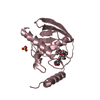
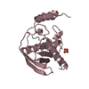
| Title | Chloro complex of the Ni-Form of E.coli deformylase | ||||||
 Components Components | PEPTIDE DEFORMYLASE | ||||||
 Keywords Keywords | HYDROLASE / PROTEIN BIOSYNTHESIS / IRON / NICKEL / METAL-BINDING | ||||||
| Function / homology |  Function and homology information Function and homology informationpeptide deformylase / peptide deformylase activity / ferrous iron binding / ribosome binding / translation / hydrolase activity / zinc ion binding / cytosol Similarity search - Function | ||||||
| Biological species |  | ||||||
| Method |  X-RAY DIFFRACTION / MOLECULAR REPLACEMENT / Resolution: 1.69 Å X-RAY DIFFRACTION / MOLECULAR REPLACEMENT / Resolution: 1.69 Å | ||||||
 Authors Authors | Ngo, Y.H.T. / Palm, G.J. / Hinrichs, W. | ||||||
 Citation Citation | Journal: J.Biol.Inorg.Chem. / Year: 2010 Title: Structure of the Ni(II) Complex of Escherichia Coli Peptide Deformylase and Suggestions on Deformylase Activities Depending on Different Metal(II) Centres. Authors: Yen, N.T.H. / Bogdanovic, X. / Palm, G.J. / Kuhl, O. / Hinrichs, W. #1: Journal: Nat.Struct.Biol. / Year: 1998Title: Iron Center, Substrate Recognition and Mechanism of Peptide Deformylase. Authors: Becker, A. / Schlichting, I. / Kabsch, W. / Groche, D. / Schultz, S. / Wagner, A.F. | ||||||
| History |
|
- Structure visualization
| Structure viewer | Molecule: MolmilJmol/JSmol |
|---|
- Downloads & links
-Download
| PDBx/mmCIF format | 2w3t.cif.gz | 53.5 KB | Display | PDBx/mmCIF format |
|---|---|---|---|---|
| PDB format | pdb2w3t.ent.gz | 36.9 KB | Display | PDB format |
| PDBx/mmJSON format | 2w3t.json.gz | Tree view | PDBx/mmJSON format | |
| Others |  Other downloads Other downloads |
-Validation report
| Arichive directory | https://data.pdbj.org/pub/pdb/validation_reports/w3/2w3tftp://data.pdbj.org/pub/pdb/validation_reports/w3/2w3t | HTTPS FTP |
|---|
-Related structure data
| Related structure data |  2w3uC  2a18S C: citing same article ( S: Starting model for refinement |
|---|---|
| Similar structure data |


-Links
 PDBj
PDBj


- Assembly
| Deposited unit | 
| ||||||||
|---|---|---|---|---|---|---|---|---|---|
| 1 |
| ||||||||
| Unit cell |
| ||||||||
| Components on special symmetry positions |
|
-Components
| #1: Protein | Mass: 21439.650 Da / Num. of mol.: 1 / Fragment: RESIDUES 2-169 Source method: isolated from a genetically manipulated source Source: (gene. exp.) |
|---|---|
| #2: Chemical | ChemComp-NI /   Mass: 58.693 Da / Num. of mol.: 1 / Source method: obtained synthetically / Formula: Ni Mass: 58.693 Da / Num. of mol.: 1 / Source method: obtained synthetically / Formula: Ni |
| #3: Chemical | ChemComp-CL /   Mass: 35.453 Da / Num. of mol.: 1 / Source method: obtained synthetically / Formula: Cl Mass: 35.453 Da / Num. of mol.: 1 / Source method: obtained synthetically / Formula: Cl |
| #4: Chemical | ChemComp-EOH /   Mass: 46.068 Da / Num. of mol.: 1 / Source method: obtained synthetically / Formula: C2H6O Mass: 46.068 Da / Num. of mol.: 1 / Source method: obtained synthetically / Formula: C2H6O |
| #5: Water | ChemComp-HOH /  Mass: 18.015 Da / Num. of mol.: 176 / Source method: isolated from a natural source / Formula: H2O Mass: 18.015 Da / Num. of mol.: 176 / Source method: isolated from a natural source / Formula: H2O |
-Experimental details
-Experiment
| Experiment | Method: X-RAY DIFFRACTION / Number of used crystals: 1 |
|---|
- Sample preparation
| Crystal | Density Matthews: 2.14 Å3/Da / Density % sol: 42.5 % / Description: NONE |
|---|---|
| Crystal grow | Temperature: 293 K / pH: 4 / Details: 20.5% PEG4000, 100MM NAOAC PH 4.0, 293 K |
-Data collection
| Diffraction | Mean temperature: 110 K |
|---|---|
| Diffraction source | Source: ROTATING ANODE / Type: RIGAKU MICROMAX-007 / Wavelength: 1.5418 |
| Detector | Type: RIGAKU CCD / Detector: CCD / Date: Apr 8, 2008 / Details: OSMIC MULTILAYER |
| Radiation | Monochromator: OSMIC MULTILAYER / Protocol: SINGLE WAVELENGTH / Monochromatic (M) / Laue (L): M / Scattering type: x-ray |
| Radiation wavelength | Wavelength: 1.5418 Å / Relative weight: 1 |
| Reflection | Resolution: 1.69→40 Å / Num. obs: 19326 / % possible obs: 94.2 % / Redundancy: 4.29 % / Biso Wilson estimate: 28 Å2 / Rmerge(I) obs: 0.05 / Net I/σ(I): 18 |
| Reflection shell | Resolution: 1.69→1.75 Å / Redundancy: 1.74 % / Rmerge(I) obs: 0.28 / Mean I/σ(I) obs: 1.8 / % possible all: 56.5 |
- Processing
| Software |
| ||||||||||||||||||||||||||||||||||||||||||||||||||||||||||||||||||||||||||||||||||||||||||||||||||||||||||||||||||||||||||||||||||||||||||||||||||||||||||||||||||||||||||||||||||||||
|---|---|---|---|---|---|---|---|---|---|---|---|---|---|---|---|---|---|---|---|---|---|---|---|---|---|---|---|---|---|---|---|---|---|---|---|---|---|---|---|---|---|---|---|---|---|---|---|---|---|---|---|---|---|---|---|---|---|---|---|---|---|---|---|---|---|---|---|---|---|---|---|---|---|---|---|---|---|---|---|---|---|---|---|---|---|---|---|---|---|---|---|---|---|---|---|---|---|---|---|---|---|---|---|---|---|---|---|---|---|---|---|---|---|---|---|---|---|---|---|---|---|---|---|---|---|---|---|---|---|---|---|---|---|---|---|---|---|---|---|---|---|---|---|---|---|---|---|---|---|---|---|---|---|---|---|---|---|---|---|---|---|---|---|---|---|---|---|---|---|---|---|---|---|---|---|---|---|---|---|---|---|---|---|
| Refinement | Method to determine structure: MOLECULAR REPLACEMENT Starting model: PDB ENTRY 2A18 Resolution: 1.69→62.02 Å / Cor.coef. Fo:Fc: 0.957 / Cor.coef. Fo:Fc free: 0.942 / SU B: 5.744 / SU ML: 0.084 / TLS residual ADP flag: LIKELY RESIDUAL / Cross valid method: THROUGHOUT / ESU R: 0.14 / ESU R Free: 0.129 / Stereochemistry target values: MAXIMUM LIKELIHOOD Details: HYDROGENS HAVE BEEN ADDED IN THE RIDING POSITIONS. ONLY RESIDUAL U VALUES ARE SHOWN. CL A 1002 AND HOH A 1004 TO 1005 ALTERNATIVELY OCCUPY THE ACTIVE SITE HOH A A 1004 AND HOH A 1005 HAVE ...Details: HYDROGENS HAVE BEEN ADDED IN THE RIDING POSITIONS. ONLY RESIDUAL U VALUES ARE SHOWN. CL A 1002 AND HOH A 1004 TO 1005 ALTERNATIVELY OCCUPY THE ACTIVE SITE HOH A A 1004 AND HOH A 1005 HAVE BEEN REFINED WITH THE DISTANCE RESTRAINED TO 2.6 A
| ||||||||||||||||||||||||||||||||||||||||||||||||||||||||||||||||||||||||||||||||||||||||||||||||||||||||||||||||||||||||||||||||||||||||||||||||||||||||||||||||||||||||||||||||||||||
| Solvent computation | Ion probe radii: 0.8 Å / Shrinkage radii: 0.8 Å / VDW probe radii: 1.2 Å / Solvent model: MASK | ||||||||||||||||||||||||||||||||||||||||||||||||||||||||||||||||||||||||||||||||||||||||||||||||||||||||||||||||||||||||||||||||||||||||||||||||||||||||||||||||||||||||||||||||||||||
| Displacement parameters | Biso mean: 18.88 Å2
| ||||||||||||||||||||||||||||||||||||||||||||||||||||||||||||||||||||||||||||||||||||||||||||||||||||||||||||||||||||||||||||||||||||||||||||||||||||||||||||||||||||||||||||||||||||||
| Refinement step | Cycle: LAST / Resolution: 1.69→62.02 Å
| ||||||||||||||||||||||||||||||||||||||||||||||||||||||||||||||||||||||||||||||||||||||||||||||||||||||||||||||||||||||||||||||||||||||||||||||||||||||||||||||||||||||||||||||||||||||
| Refine LS restraints |
|