 Movie
Movie Controller

[English] 日本語
 Yorodumi
- PDB-1lru: Crystal Structure of E.coli Peptide Deformylase Complexed with An... -
+ Open data

- Basic information
| Entry | Database: PDB / ID: 1lru | ||||||
|---|---|---|---|---|---|---|---|
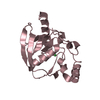
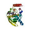
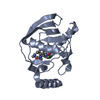
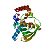
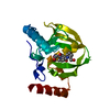
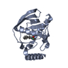
| Title | Crystal Structure of E.coli Peptide Deformylase Complexed with Antibiotic Actinonin | ||||||
 Components Components | PEPTIDE DEFORMYLASE | ||||||
 Keywords Keywords | HYDROLASE / ACTINONIN / INHIBITION / POLYPEPTIDE DEFORMYLASE | ||||||
| Function / homology |  Function and homology information Function and homology informationpeptide deformylase / peptide deformylase activity / ferrous iron binding / ribosome binding / translation / hydrolase activity / zinc ion binding / cytosol Similarity search - Function | ||||||
| Biological species |  | ||||||
| Method |  X-RAY DIFFRACTION / SIRAS / Resolution: 2.1 Å X-RAY DIFFRACTION / SIRAS / Resolution: 2.1 Å | ||||||
 Authors Authors | Guilloteau, J.-P. / Mathieu, M. / Giglione, C. / Blanc, V. / Dupuy, A. / Chevrier, M. / Gil, P. / Famechon, A. / Meinnel, T. / Mikol, V. | ||||||
 Citation Citation | Journal: J.Mol.Biol. / Year: 2002 Title: The crystal structures of four peptide deformylases bound to the antibiotic actinonin reveal two distinct types: a platform for the structure-based design of antibacterial agents. Authors: Guilloteau, J.P. / Mathieu, M. / Giglione, C. / Blanc, V. / Dupuy, A. / Chevrier, M. / Gil, P. / Famechon, A. / Meinnel, T. / Mikol, V. | ||||||
| History |
|
- Structure visualization
| Structure viewer | Molecule: MolmilJmol/JSmol |
|---|
- Downloads & links
-Download
| PDBx/mmCIF format | 1lru.cif.gz | 122.6 KB | Display | PDBx/mmCIF format |
|---|---|---|---|---|
| PDB format | pdb1lru.ent.gz | 95.3 KB | Display | PDB format |
| PDBx/mmJSON format | 1lru.json.gz | Tree view | PDBx/mmJSON format | |
| Others |  Other downloads Other downloads |
-Validation report
| Arichive directory | https://data.pdbj.org/pub/pdb/validation_reports/lr/1lruftp://data.pdbj.org/pub/pdb/validation_reports/lr/1lru | HTTPS FTP |
|---|
-Related structure data


-Links
 PDBj
PDBj- Assembly
| Deposited unit | 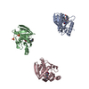
| ||||||||
|---|---|---|---|---|---|---|---|---|---|
| 1 | 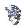
| ||||||||
| 2 | 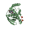
| ||||||||
| 3 | 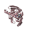
| ||||||||
| 4 | 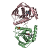
| ||||||||
| Unit cell |
| ||||||||
| Details | the biological unit is the monomer. There are three monomers in the asymetric unit. |
-Components
| #1: Protein | Mass: 19226.248 Da / Num. of mol.: 3 Source method: isolated from a genetically manipulated source Source: (gene. exp.) #2: Chemical |   Mass: 65.409 Da / Num. of mol.: 3 / Source method: obtained synthetically / Formula: Zn Mass: 65.409 Da / Num. of mol.: 3 / Source method: obtained synthetically / Formula: Zn#3: Chemical |   Mass: 385.498 Da / Num. of mol.: 3 / Source method: obtained synthetically / Formula: C19H35N3O5 / Comment: antitumor, antibiotic*YM Mass: 385.498 Da / Num. of mol.: 3 / Source method: obtained synthetically / Formula: C19H35N3O5 / Comment: antitumor, antibiotic*YM#4: Chemical |   Mass: 96.063 Da / Num. of mol.: 2 / Source method: obtained synthetically / Formula: SO4 Mass: 96.063 Da / Num. of mol.: 2 / Source method: obtained synthetically / Formula: SO4#5: Water | ChemComp-HOH / |  Mass: 18.015 Da / Num. of mol.: 486 / Source method: isolated from a natural source / Formula: H2O Mass: 18.015 Da / Num. of mol.: 486 / Source method: isolated from a natural source / Formula: H2O |
|---|
-Experimental details
-Experiment
| Experiment | Method: X-RAY DIFFRACTION / Number of used crystals: 1 |
|---|
- Sample preparation
| Crystal | Density Matthews: 2.6 Å3/Da / Density % sol: 54.63 % | ||||||||||||||||||||||||||||||||||||||||||
|---|---|---|---|---|---|---|---|---|---|---|---|---|---|---|---|---|---|---|---|---|---|---|---|---|---|---|---|---|---|---|---|---|---|---|---|---|---|---|---|---|---|---|---|
| Crystal grow | Temperature: 290 K / Method: vapor diffusion, hanging drop / pH: 6 Details: 0.5M (NH4)2SO4, 28%PEG400, pH 6.0, VAPOR DIFFUSION, HANGING DROP, temperature 290K | ||||||||||||||||||||||||||||||||||||||||||
| Crystal grow | *PLUS Temperature: 19 ℃ | ||||||||||||||||||||||||||||||||||||||||||
| Components of the solutions | *PLUS
|
-Data collection
| Diffraction | Mean temperature: 95 K |
|---|---|
| Diffraction source | Source: ROTATING ANODE / Type: ENRAF-NONIUS / Wavelength: 1.5418 Å |
| Detector | Type: MAC Science DIP-2000 / Detector: IMAGE PLATE / Date: Apr 14, 1997 |
| Radiation | Monochromator: Ni MIRROR + Ni FILTER / Protocol: SINGLE WAVELENGTH / Monochromatic (M) / Laue (L): M / Scattering type: x-ray |
| Radiation wavelength | Wavelength: 1.5418 Å / Relative weight: 1 |
| Reflection | Resolution: 2.1→15 Å / Num. all: 34394 / Num. obs: 34394 / % possible obs: 93.7 % / Observed criterion σ(F): 0 / Observed criterion σ(I): 0 / Redundancy: 2.4 % / Rsym value: 0.076 |
| Reflection shell | Resolution: 2.1→2.17 Å / Redundancy: 2.3 % / Num. unique all: 3037 / Rsym value: 0.251 / % possible all: 83.7 |
| Reflection | *PLUS Lowest resolution: 15 Å / Rmerge(I) obs: 0.076 |
| Reflection shell | *PLUS Highest resolution: 2.1 Å / % possible obs: 83.7 % / Rmerge(I) obs: 0.251 / Mean I/σ(I) obs: 5 |
- Processing
| Software |
| ||||||||||||
|---|---|---|---|---|---|---|---|---|---|---|---|---|---|
| Refinement | Method to determine structure: SIRAS / Resolution: 2.1→15 Å / σ(F): 0 / Stereochemistry target values: Engh & Huber
| ||||||||||||
| Refinement step | Cycle: LAST / Resolution: 2.1→15 Å
| ||||||||||||
| Refine LS restraints |
| ||||||||||||
| Refinement | *PLUS Highest resolution: 2.1 Å / Rfactor obs: 0.24 / Rfactor Rwork: 0.24 | ||||||||||||
| Solvent computation | *PLUS | ||||||||||||
| Displacement parameters | *PLUS |
