 Movie
Movie Controller


+ Open data

- Basic information
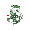
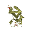
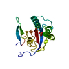
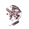
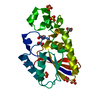
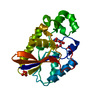
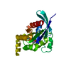
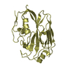
| Entry | Database: PDB / ID: 1bs5 | ||||||
|---|---|---|---|---|---|---|---|
| Title | PEPTIDE DEFORMYLASE AS ZN2+ CONTAINING FORM | ||||||
 Components Components | PROTEIN (PEPTIDE DEFORMYLASE) | ||||||
 Keywords Keywords | HYDROLASE / IRON METALLOPROTEASE / PROTEIN SYNTHESIS | ||||||
| Function / homology |  Function and homology information Function and homology informationpeptide deformylase / peptide deformylase activity / ferrous iron binding / ribosome binding / translation / hydrolase activity / zinc ion binding / cytosol Similarity search - Function | ||||||
| Biological species |  | ||||||
| Method |  X-RAY DIFFRACTION / MOLECULAR REPLACEMENT / Resolution: 2.5 Å X-RAY DIFFRACTION / MOLECULAR REPLACEMENT / Resolution: 2.5 Å | ||||||
 Authors Authors | Becker, A. / Schlichting, I. / Kabsch, W. / Groche, D. / Schultz, S. / Wagner, A.F.V. | ||||||
 Citation Citation | Journal: Nat.Struct.Biol. / Year: 1998 Title: Iron center, substrate recognition and mechanism of peptide deformylase. Authors: Becker, A. / Schlichting, I. / Kabsch, W. / Groche, D. / Schultz, S. / Wagner, A.F. #1: Journal: J.Biol.Chem. / Year: 1998Title: Structure of Peptide Deformylase and Identification of the Substrate Binding Site Authors: Becker, A. / Schlichting, I. / Kabsch, W. / Schultz, S. / Wagner, A.F.V. #2: Journal: Biochem.Biophys.Res.Commun. / Year: 1998Title: Isolation and Crystallization of Functionally Competent Escherichia Coli Peptide Deformylase Forms Containing Either Iron or Nickel in the Active Site Authors: Groche, D. / Becker, A. / Schlichting, I. / Kabsch, W. / Schultz, S. / Wagner, A.F.V. | ||||||
| History |
|
- Structure visualization
| Structure viewer | Molecule: MolmilJmol/JSmol |
|---|
- Downloads & links
-Download
| PDBx/mmCIF format | 1bs5.cif.gz | 114.4 KB | Display | PDBx/mmCIF format |
|---|---|---|---|---|
| PDB format | pdb1bs5.ent.gz | 88 KB | Display | PDB format |
| PDBx/mmJSON format | 1bs5.json.gz | Tree view | PDBx/mmJSON format | |
| Others |  Other downloads Other downloads |
-Validation report
| Arichive directory | https://data.pdbj.org/pub/pdb/validation_reports/bs/1bs5ftp://data.pdbj.org/pub/pdb/validation_reports/bs/1bs5 | HTTPS FTP |
|---|
-Related structure data
| Related structure data | 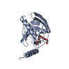 1bs4C  1bs6C  1bs8C  1bszC  1icjS C: citing same article ( S: Starting model for refinement |
|---|---|
| Similar structure data |


-Links
 PDBj
PDBj- Assembly
| Deposited unit | 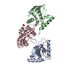
| ||||||||
|---|---|---|---|---|---|---|---|---|---|
| 1 | 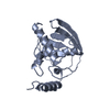
| ||||||||
| 2 | 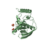
| ||||||||
| 3 | 
| ||||||||
| Unit cell |
|
-Components
| #1: Protein | Mass: 19226.248 Da / Num. of mol.: 3 Source method: isolated from a genetically manipulated source Details: PDF PROTEIN FROM ESCHERICHIA COLI IS CRYSTALLIZED AS ZN2+ CONTAINING FORM Source: (gene. exp.) #2: Chemical |   Mass: 65.409 Da / Num. of mol.: 3 / Source method: obtained synthetically / Formula: Zn Mass: 65.409 Da / Num. of mol.: 3 / Source method: obtained synthetically / Formula: Zn#3: Chemical |   Mass: 96.063 Da / Num. of mol.: 2 / Source method: obtained synthetically / Formula: SO4 Mass: 96.063 Da / Num. of mol.: 2 / Source method: obtained synthetically / Formula: SO4#4: Water | ChemComp-HOH / |  Mass: 18.015 Da / Num. of mol.: 132 / Source method: isolated from a natural source / Formula: H2O Mass: 18.015 Da / Num. of mol.: 132 / Source method: isolated from a natural source / Formula: H2O |
|---|
-Experimental details
-Experiment
| Experiment | Method: X-RAY DIFFRACTION / Number of used crystals: 5 |
|---|
- Sample preparation
| Crystal | Density Matthews: 2.82 Å3/Da / Density % sol: 56 % | ||||||||||||||||||||||||||||||
|---|---|---|---|---|---|---|---|---|---|---|---|---|---|---|---|---|---|---|---|---|---|---|---|---|---|---|---|---|---|---|---|
| Crystal grow | pH: 7.4 Details: SEE: D.GROCHE,A.BECKER,I.SCHLICHTING,W.KABSCH, S.SCHULTZ,A.F.V.WAGNER (1998) BIOCHEM.BIOPHYS.RES.COMM. 246, 342, pH 7.4 | ||||||||||||||||||||||||||||||
| Crystal | *PLUS | ||||||||||||||||||||||||||||||
| Crystal grow | *PLUS Method: vapor diffusion, hanging dropDetails: Groche, D., (1998) Biochem.Biophys.Res.Comm., 246, 342. | ||||||||||||||||||||||||||||||
| Components of the solutions | *PLUS
|
-Data collection
| Diffraction | Mean temperature: 293 K |
|---|---|
| Diffraction source | Source: ROTATING ANODE / Type: ELLIOTT GX-18 / Wavelength: 1.5418 |
| Detector | Type: SIEMENS-NICOLET X100 / Detector: AREA DETECTOR / Date: Jul 15, 1997 / Details: FRANCKS DUBBLE-MIRROR OPTICS |
| Radiation | Monochromator: NI FILTER / Protocol: SINGLE WAVELENGTH / Monochromatic (M) / Laue (L): M / Scattering type: x-ray |
| Radiation wavelength | Wavelength: 1.5418 Å / Relative weight: 1 |
| Reflection | Resolution: 2.5→20 Å / Num. obs: 22399 / % possible obs: 99.3 % / Redundancy: 5.8 % / Biso Wilson estimate: 43.6 Å2 / Rmerge(I) obs: 0.076 / Net I/σ(I): 15.72 |
| Reflection shell | Resolution: 2.5→2.64 Å / Redundancy: 2.8 % / Rmerge(I) obs: 0.338 / Mean I/σ(I) obs: 1.9 / % possible all: 99.4 |
| Reflection | *PLUS Num. measured all: 130772 |
- Processing
| Software |
| ||||||||||||||||||||||||||||||||||||||||||||||||||||||||||||||||||||||||||||||||
|---|---|---|---|---|---|---|---|---|---|---|---|---|---|---|---|---|---|---|---|---|---|---|---|---|---|---|---|---|---|---|---|---|---|---|---|---|---|---|---|---|---|---|---|---|---|---|---|---|---|---|---|---|---|---|---|---|---|---|---|---|---|---|---|---|---|---|---|---|---|---|---|---|---|---|---|---|---|---|---|---|---|
| Refinement | Method to determine structure: MOLECULAR REPLACEMENT Starting model: 1ICJ Resolution: 2.5→6 Å / Rfactor Rfree error: 0.006 / Data cutoff high absF: 100000 / Data cutoff low absF: 0 / Isotropic thermal model: RESTRAINED / Cross valid method: THROUGHOUT / σ(F): 0 / Details: BULK SOLVENT MODEL USED
| ||||||||||||||||||||||||||||||||||||||||||||||||||||||||||||||||||||||||||||||||
| Displacement parameters | Biso mean: 37 Å2
| ||||||||||||||||||||||||||||||||||||||||||||||||||||||||||||||||||||||||||||||||
| Refine analyze |
| ||||||||||||||||||||||||||||||||||||||||||||||||||||||||||||||||||||||||||||||||
| Refinement step | Cycle: LAST / Resolution: 2.5→6 Å
| ||||||||||||||||||||||||||||||||||||||||||||||||||||||||||||||||||||||||||||||||
| Refine LS restraints |
| ||||||||||||||||||||||||||||||||||||||||||||||||||||||||||||||||||||||||||||||||
| LS refinement shell | Resolution: 2.5→2.64 Å / Rfactor Rfree error: 0.02 / Total num. of bins used: 6
| ||||||||||||||||||||||||||||||||||||||||||||||||||||||||||||||||||||||||||||||||
| Xplor file |
| ||||||||||||||||||||||||||||||||||||||||||||||||||||||||||||||||||||||||||||||||
| Software | *PLUS Name: X-PLOR / Version: 3.851 / Classification: refinement | ||||||||||||||||||||||||||||||||||||||||||||||||||||||||||||||||||||||||||||||||
| Refinement | *PLUS Lowest resolution: 6 Å / σ(F): 0 / % reflection Rfree: 10.4 % | ||||||||||||||||||||||||||||||||||||||||||||||||||||||||||||||||||||||||||||||||
| Solvent computation | *PLUS | ||||||||||||||||||||||||||||||||||||||||||||||||||||||||||||||||||||||||||||||||
| Displacement parameters | *PLUS Biso mean: 37 Å2 | ||||||||||||||||||||||||||||||||||||||||||||||||||||||||||||||||||||||||||||||||
| Refine LS restraints | *PLUS
| ||||||||||||||||||||||||||||||||||||||||||||||||||||||||||||||||||||||||||||||||
| LS refinement shell | *PLUS Rfactor Rfree: 0.349 / % reflection Rfree: 9.2 % / Rfactor Rwork: 0.294 |
