 Movie
Movie Controller

[English] 日本語
 Yorodumi
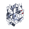
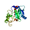
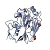
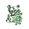
- PDB-1h2f: BACILLUS STEAROTHERMOPHILUS PHOE (previously known as yhfr) in co... -
+ Open data

- Basic information
| Entry | Database: PDB / ID: 1h2f | ||||||
|---|---|---|---|---|---|---|---|
| Title | BACILLUS STEAROTHERMOPHILUS PHOE (previously known as yhfr) in complex with trivanadate | ||||||
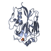
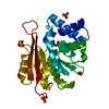
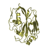
 Components Components | PHOSPHATASE | ||||||
 Keywords Keywords | HYDROLASE / BROAD SPECIFICITY PHOSPHATASE / DPGM HOMOLOG | ||||||
| Function / homology |  Function and homology information Function and homology information | ||||||
| Biological species |   BACILLUS STEAROTHERMOPHILUS (bacteria) BACILLUS STEAROTHERMOPHILUS (bacteria) | ||||||
| Method |  X-RAY DIFFRACTION / SYNCHROTRON / OTHER / Resolution: 2 Å X-RAY DIFFRACTION / SYNCHROTRON / OTHER / Resolution: 2 Å | ||||||
 Authors Authors | Rigden, D.J. / Littlejohn, J.E. / Jedrzejas, M.J. | ||||||
 Citation Citation | Journal: J.Mol.Biol. / Year: 2003 Title: Structures of Phosphate and Trivanadate Complexes of Bacillus Stearothermophilus Phosphatase Phoe: Structural and Functional Analysis in the Cofactor-Dependent Phosphoglycerate Mutase Superfamily Authors: Rigden, D.J. / Littlejohn, J.E. / Henderson, K. / Jedrzejas, M.J. #1: Journal: J.Mol.Biol. / Year: 2002Title: Structure and Mechanism of Action of a Cofactor-Dependent Phosphoglycerate Mutase Homolog from Bacillus Stearothermophilus with Broad Specificity Phosphatase Activity Authors: Rigden, D.J. / Mello, L.V. / Setlow, P. / Jedrzejas, M.J. #2: Journal: Protein Sci. / Year: 2001 Title: A Cofactor-Dependent Phosphoglycerate Mutase Homolog from Bacillus Stearothermophilus is Actually a Broad Specificity Phosphatase Authors: Rigden, D.J. / Bagyan, I. / Lamani, E. / Setlow, P. / Jedrzejas, M.J. | ||||||
| History |
|
- Structure visualization
| Structure viewer | Molecule: MolmilJmol/JSmol |
|---|
- Downloads & links
-Download
| PDBx/mmCIF format | 1h2f.cif.gz | 60.1 KB | Display | PDBx/mmCIF format |
|---|---|---|---|---|
| PDB format | pdb1h2f.ent.gz | 44.2 KB | Display | PDB format |
| PDBx/mmJSON format | 1h2f.json.gz | Tree view | PDBx/mmJSON format | |
| Others |  Other downloads Other downloads |
-Validation report
| Arichive directory | https://data.pdbj.org/pub/pdb/validation_reports/h2/1h2fftp://data.pdbj.org/pub/pdb/validation_reports/h2/1h2f | HTTPS FTP |
|---|
-Related structure data


-Links
 PDBj
PDBj


- Assembly
| Deposited unit | 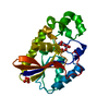
| ||||||||
|---|---|---|---|---|---|---|---|---|---|
| 1 |
| ||||||||
| Unit cell |
|
-Components
| #1: Protein | Mass: 23647.676 Da / Num. of mol.: 1 Source method: isolated from a genetically manipulated source Source: (gene. exp.) BACILLUS STEAROTHERMOPHILUS (bacteria) / Plasmid: PS3297 / Production host: |
|---|---|
| #2: Chemical | ChemComp-VA3 /   Mass: 280.820 Da / Num. of mol.: 1 / Source method: obtained synthetically / Formula: O8V3 Mass: 280.820 Da / Num. of mol.: 1 / Source method: obtained synthetically / Formula: O8V3 |
| #3: Chemical | ChemComp-PO4 /   Mass: 94.971 Da / Num. of mol.: 1 / Source method: obtained synthetically / Formula: PO4 Mass: 94.971 Da / Num. of mol.: 1 / Source method: obtained synthetically / Formula: PO4 |
| #4: Water | ChemComp-HOH /  Mass: 18.015 Da / Num. of mol.: 142 / Source method: isolated from a natural source / Formula: H2O Mass: 18.015 Da / Num. of mol.: 142 / Source method: isolated from a natural source / Formula: H2O |
| Compound details | THE SWISSPROT ENTRY Q9ALU0 IDENTIFIES THIS PROTEIN AS A PHOSPHOGLYCERATE MUTASE. THE PROTEIN ...THE SWISSPROT ENTRY Q9ALU0 IDENTIFIES |
| Has protein modification | Y |
| Sequence details | THE SWISSPROT ENTRY Q9ALU0 IDENTIFIES THIS PROTEIN AS A PHOSPHOGLYCERATE MUTASE. THE PROTEIN ...THE SWISSPROT ENTRY Q9ALU0 IDENTIFIES |
-Experimental details
-Experiment
| Experiment | Method: X-RAY DIFFRACTION / Number of used crystals: 1 |
|---|
- Sample preparation
| Crystal | Density Matthews: 2.27 Å3/Da / Density % sol: 45.3 % | |||||||||||||||||||||||||||||||||||||||||||||||||
|---|---|---|---|---|---|---|---|---|---|---|---|---|---|---|---|---|---|---|---|---|---|---|---|---|---|---|---|---|---|---|---|---|---|---|---|---|---|---|---|---|---|---|---|---|---|---|---|---|---|---|
| Crystal grow | pH: 4.5 Details: 15 % ETHYLENE GLYCOL, 85 MM SODIUM CACODYLATE PH 4.5 | |||||||||||||||||||||||||||||||||||||||||||||||||
| Crystal grow | *PLUS Temperature: 22 ℃ / pH: 7.4 / Method: vapor diffusion, hanging drop | |||||||||||||||||||||||||||||||||||||||||||||||||
| Components of the solutions | *PLUS
|
-Data collection
| Diffraction | Mean temperature: 100 K |
|---|---|
| Diffraction source | Source: SYNCHROTRON / Site: ALS  / Beamline: 5.0.1 / Wavelength: 1 / Beamline: 5.0.1 / Wavelength: 1 |
| Detector | Type: ADSC CCD / Detector: CCD / Date: Mar 15, 2002 |
| Radiation | Protocol: SINGLE WAVELENGTH / Monochromatic (M) / Laue (L): M / Scattering type: x-ray |
| Radiation wavelength | Wavelength: 1 Å / Relative weight: 1 |
| Reflection | Resolution: 2→50 Å / Num. obs: 17334 / % possible obs: 95.2 % / Redundancy: 4.9 % / Biso Wilson estimate: 29.6 Å2 / Rmerge(I) obs: 0.155 / Net I/σ(I): 15.8 |
| Reflection shell | Resolution: 2→2.07 Å / Redundancy: 2.1 % / Rmerge(I) obs: 0.554 / Mean I/σ(I) obs: 2.1 / % possible all: 88.2 |
| Reflection | *PLUS Lowest resolution: 50 Å |
| Reflection shell | *PLUS % possible obs: 88.2 % / Mean I/σ(I) obs: 1.4 |
- Processing
| Software |
| ||||||||||||||||||||||||||||||||||||||||||||||||||||||||||||||||||||||||||||||||
|---|---|---|---|---|---|---|---|---|---|---|---|---|---|---|---|---|---|---|---|---|---|---|---|---|---|---|---|---|---|---|---|---|---|---|---|---|---|---|---|---|---|---|---|---|---|---|---|---|---|---|---|---|---|---|---|---|---|---|---|---|---|---|---|---|---|---|---|---|---|---|---|---|---|---|---|---|---|---|---|---|---|
| Refinement | Method to determine structure: OTHER / Resolution: 2→50 Å / Rfactor Rfree error: 0.008 / Data cutoff high absF: 1636651.82 / Isotropic thermal model: RESTRAINED / Cross valid method: THROUGHOUT / σ(F): 0 / Stereochemistry target values: MAXIMUM LIKELIHOOD Details: CRYSTAL WAS EXPOSED TO 50MM AMMONIUM VANADATE, 30% ETHYLENE GLYCOL, 20MM SODIUM ACETATE PH 5.0 FOR 3 DAYS BEFORE DATA COLLECTION.
| ||||||||||||||||||||||||||||||||||||||||||||||||||||||||||||||||||||||||||||||||
| Solvent computation | Solvent model: FLAT MODEL / Bsol: 45.1339 Å2 / ksol: 0.307577 e/Å3 | ||||||||||||||||||||||||||||||||||||||||||||||||||||||||||||||||||||||||||||||||
| Displacement parameters | Biso mean: 42.7 Å2
| ||||||||||||||||||||||||||||||||||||||||||||||||||||||||||||||||||||||||||||||||
| Refine analyze |
| ||||||||||||||||||||||||||||||||||||||||||||||||||||||||||||||||||||||||||||||||
| Refinement step | Cycle: LAST / Resolution: 2→50 Å
| ||||||||||||||||||||||||||||||||||||||||||||||||||||||||||||||||||||||||||||||||
| Refine LS restraints |
| ||||||||||||||||||||||||||||||||||||||||||||||||||||||||||||||||||||||||||||||||
| LS refinement shell | Resolution: 2→2.07 Å / Rfactor Rfree error: 0.042 / Total num. of bins used: 10
| ||||||||||||||||||||||||||||||||||||||||||||||||||||||||||||||||||||||||||||||||
| Xplor file |
| ||||||||||||||||||||||||||||||||||||||||||||||||||||||||||||||||||||||||||||||||
| Refinement | *PLUS Lowest resolution: 50 Å | ||||||||||||||||||||||||||||||||||||||||||||||||||||||||||||||||||||||||||||||||
| Solvent computation | *PLUS | ||||||||||||||||||||||||||||||||||||||||||||||||||||||||||||||||||||||||||||||||
| Displacement parameters | *PLUS | ||||||||||||||||||||||||||||||||||||||||||||||||||||||||||||||||||||||||||||||||
| Refine LS restraints | *PLUS
| ||||||||||||||||||||||||||||||||||||||||||||||||||||||||||||||||||||||||||||||||
| LS refinement shell | *PLUS Rfactor Rfree: 0.358 / Rfactor Rwork: 0.338 / Num. reflection Rwork: 1942 |
