 Movie
Movie Controller

[English] 日本語
 Yorodumi
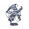
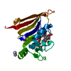
- PDB-1g2a: THE CRYSTAL STRUCTURE OF E.COLI PEPTIDE DEFORMYLASE COMPLEXED WIT... -
+ Open data

- Basic information
| Entry | Database: PDB / ID: 1g2a | ||||||
|---|---|---|---|---|---|---|---|
| Title | THE CRYSTAL STRUCTURE OF E.COLI PEPTIDE DEFORMYLASE COMPLEXED WITH ACTINONIN | ||||||
 Components Components | POLYPEPTIDE DEFORMYLASE | ||||||
 Keywords Keywords | HYDROLASE / actinonin / inhibition / Polypeptide deformylase | ||||||
| Function / homology |  Function and homology information Function and homology informationpeptide deformylase / peptide deformylase activity / ferrous iron binding / ribosome binding / translation / hydrolase activity / zinc ion binding / cytosol Similarity search - Function | ||||||
| Biological species |  | ||||||
| Method |  X-RAY DIFFRACTION / MOLECULAR REPLACEMENT / Resolution: 1.75 Å X-RAY DIFFRACTION / MOLECULAR REPLACEMENT / Resolution: 1.75 Å | ||||||
 Authors Authors | Clements, J.M. / Beckett, P. / Brown, A. / Catlin, C. / Lobell, M. / Palan, S. / Thomas, W. / Whittaker, M. / Baker, P.J. / Rodgers, H.F. ...Clements, J.M. / Beckett, P. / Brown, A. / Catlin, C. / Lobell, M. / Palan, S. / Thomas, W. / Whittaker, M. / Baker, P.J. / Rodgers, H.F. / Barynin, V. / Rice, D.W. / Hunter, M.G. | ||||||
 Citation Citation | Journal: Antimicrob.Agents Chemother. / Year: 2001 Title: Antibiotic activity and characterization of BB-3497, a novel peptide deformylase inhibitor. Authors: Clements, J.M. / Beckett, R.P. / Brown, A. / Catlin, G. / Lobell, M. / Palan, S. / Thomas, W. / Whittaker, M. / Wood, S. / Salama, S. / Baker, P.J. / Rodgers, H.F. / Barynin, V. / Rice, D.W. / Hunter, M.G. | ||||||
| History |
|
- Structure visualization
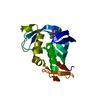
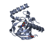
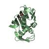
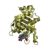
| Structure viewer | Molecule: MolmilJmol/JSmol |
|---|
- Downloads & links
-Download
| PDBx/mmCIF format | 1g2a.cif.gz | 129.8 KB | Display | PDBx/mmCIF format |
|---|---|---|---|---|
| PDB format | pdb1g2a.ent.gz | 100.2 KB | Display | PDB format |
| PDBx/mmJSON format | 1g2a.json.gz | Tree view | PDBx/mmJSON format | |
| Others |  Other downloads Other downloads |
-Validation report
| Arichive directory | https://data.pdbj.org/pub/pdb/validation_reports/g2/1g2aftp://data.pdbj.org/pub/pdb/validation_reports/g2/1g2a | HTTPS FTP |
|---|
-Related structure data


-Links
 PDBj
PDBj- Assembly
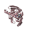
| Deposited unit | 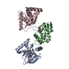
| ||||||||||
|---|---|---|---|---|---|---|---|---|---|---|---|
| 1 | 
| ||||||||||
| 2 | 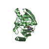
| ||||||||||
| 3 | 
| ||||||||||
| Unit cell |
|
-Components
| #1: Protein | Mass: 19226.248 Da / Num. of mol.: 3 Source method: isolated from a genetically manipulated source Source: (gene. exp.) #2: Chemical |   Mass: 58.693 Da / Num. of mol.: 3 / Source method: obtained synthetically / Formula: Ni Mass: 58.693 Da / Num. of mol.: 3 / Source method: obtained synthetically / Formula: Ni#3: Chemical |   Mass: 385.498 Da / Num. of mol.: 3 / Source method: obtained synthetically / Formula: C19H35N3O5 / Comment: antitumor, antibiotic*YM Mass: 385.498 Da / Num. of mol.: 3 / Source method: obtained synthetically / Formula: C19H35N3O5 / Comment: antitumor, antibiotic*YM#4: Water | ChemComp-HOH / |  Mass: 18.015 Da / Num. of mol.: 725 / Source method: isolated from a natural source / Formula: H2O Mass: 18.015 Da / Num. of mol.: 725 / Source method: isolated from a natural source / Formula: H2O |
|---|
-Experimental details
-Experiment
| Experiment | Method: X-RAY DIFFRACTION / Number of used crystals: 1 |
|---|
- Sample preparation
| Crystal | Density Matthews: 2.77 Å3/Da / Density % sol: 55.6 % | |||||||||||||||||||||||||||||||||||
|---|---|---|---|---|---|---|---|---|---|---|---|---|---|---|---|---|---|---|---|---|---|---|---|---|---|---|---|---|---|---|---|---|---|---|---|---|
| Crystal grow | Temperature: 290 K / Method: vapor diffusion, hanging drop / pH: 7.5 Details: 10mg/ml PDF, 20mM actinonin, 50mM HEPES, pH 7.5 + 25-32% PEG 4000, 0.1M sodium citrate, pH 5.6, 0.2M ammonium acetate, VAPOR DIFFUSION, HANGING DROP at 290K | |||||||||||||||||||||||||||||||||||
| Crystal grow | *PLUS | |||||||||||||||||||||||||||||||||||
| Components of the solutions | *PLUS
|
-Data collection
| Diffraction | Mean temperature: 100 K |
|---|---|
| Diffraction source | Source: ROTATING ANODE / Type: RIGAKU RU200 / Wavelength: 1.5418 Å |
| Detector | Type: MARRESEARCH / Detector: AREA DETECTOR / Date: Apr 28, 1999 / Details: MSC Yale Mirrors |
| Radiation | Monochromator: msc yale mirrors / Protocol: SINGLE WAVELENGTH / Monochromatic (M) / Laue (L): M / Scattering type: x-ray |
| Radiation wavelength | Wavelength: 1.5418 Å / Relative weight: 1 |
| Reflection | Resolution: 1.75→14 Å / Num. all: 62907 / Num. obs: 54729 / % possible obs: 0.87 % / Observed criterion σ(F): 0 / Observed criterion σ(I): 0 / Redundancy: 1.95 % / Rmerge(I) obs: 0.044 / Net I/σ(I): 13.6 |
| Reflection shell | Resolution: 1.75→1.79 Å / Redundancy: 1.5 % / Rmerge(I) obs: 0.191 / % possible all: 86.8 |
| Reflection | *PLUS % possible obs: 85.9 % |
| Reflection shell | *PLUS % possible obs: 86.8 % / Num. unique obs: 3634 / Mean I/σ(I) obs: 3.9 |
- Processing
| Software |
| |||||||||||||||||||||||||
|---|---|---|---|---|---|---|---|---|---|---|---|---|---|---|---|---|---|---|---|---|---|---|---|---|---|---|
| Refinement | Method to determine structure: MOLECULAR REPLACEMENT / Resolution: 1.75→14 Å / Cross valid method: THROUGHOUT / σ(F): 0 / σ(I): 0 / Stereochemistry target values: Engh & Huber
| |||||||||||||||||||||||||
| Refinement step | Cycle: LAST / Resolution: 1.75→14 Å
| |||||||||||||||||||||||||
| Refine LS restraints |
| |||||||||||||||||||||||||
| Software | *PLUS Name: REFMAC / Classification: refinement | |||||||||||||||||||||||||
| Refinement | *PLUS σ(F): 0 / % reflection Rfree: 5 % / Rfactor Rfree: 0.25 / Rfactor Rwork: 0.19 | |||||||||||||||||||||||||
| Solvent computation | *PLUS | |||||||||||||||||||||||||
| Displacement parameters | *PLUS | |||||||||||||||||||||||||
| Refine LS restraints | *PLUS
|
