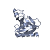
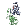
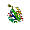
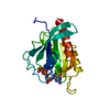
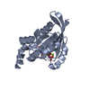
SIGNALING PROTEIN / PHOSPHOLIPASE C / PHOSPHOINOSITIDES / RHO GTPASES / RAC SIGNALING PROTEIN
Function / homology
Function and homology information
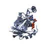
regulation of mast cell chemotaxis / regulation of cell-substrate adhesion / lymphocyte aggregation / positive regulation of mast cell proliferation / regulation of respiratory burst / regulation of mast cell degranulation / mast cell proliferation / erythrocyte enucleation / regulation of neutrophil migration / NADPH oxidase complex ...regulation of mast cell chemotaxis / regulation of cell-substrate adhesion / lymphocyte aggregation / positive regulation of mast cell proliferation / regulation of respiratory burst / regulation of mast cell degranulation / mast cell proliferation / erythrocyte enucleation / regulation of neutrophil migration / NADPH oxidase complex / regulation of hydrogen peroxide metabolic process / respiratory burst / cortical cytoskeleton organization / cell projection assembly / protein kinase regulator activity / ROS and RNS production in phagocytes / PCP/CE pathway / positive regulation of neutrophil chemotaxis / superoxide anion generation / : / small GTPase-mediated signal transduction / regulation of T cell proliferation / establishment or maintenance of cell polarity / RAC2 GTPase cycle / RHO GTPases Activate NADPH Oxidases / bone resorption / GPVI-mediated activation cascade / positive regulation of lamellipodium assembly / cell projection / actin filament organization / small monomeric GTPase / regulation of actin cytoskeleton organization / phagocytic vesicle membrane / chemotaxis / Constitutive Signaling by Aberrant PI3K in Cancer / PIP3 activates AKT signaling / nuclear envelope / regulation of cell shape / lamellipodium / PI5P, PP2A and IER3 Regulate PI3K/AKT Signaling / G protein activity / cytoplasmic vesicle / cytoskeleton / mitochondrial outer membrane / G protein-coupled receptor signaling pathway / focal adhesion / GTPase activity / protein kinase binding / endoplasmic reticulum membrane / GTP binding / signal transduction / extracellular exosome / plasma membrane / cytosol Similarity search - Function
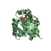
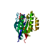
Small GTPase Rho / Small GTPase Rho domain profile. / Ran (Ras-related nuclear proteins) /TC4 subfamily of small GTPases / Rho (Ras homology) subfamily of Ras-like small GTPases / Ras subfamily of RAS small GTPases / Small GTPase / Ras family / Rab subfamily of small GTPases / Small GTP-binding protein domain / P-loop containing nucleotide triphosphate hydrolases ...Small GTPase Rho / Small GTPase Rho domain profile. / Ran (Ras-related nuclear proteins) /TC4 subfamily of small GTPases / Rho (Ras homology) subfamily of Ras-like small GTPases / Ras subfamily of RAS small GTPases / Small GTPase / Ras family / Rab subfamily of small GTPases / Small GTP-binding protein domain / P-loop containing nucleotide triphosphate hydrolases / Rossmann fold / P-loop containing nucleoside triphosphate hydrolase / 3-Layer(aba) Sandwich / Alpha Beta Similarity search - Domain/homology
Mass: 18.015 Da / Num. of mol.: 137 / Source method: isolated from a natural source / Formula: H2O
Compound details
ENGINEERED RESIDUE IN CHAIN A, GLY 12 TO VAL
Sequence details
THERE IS A LEADING LINKER SEQUENCE GGGSGGS (NOT SEEN IN STRUCTURE) AND MUTATION G12V
-
Experimental details
-
Experiment
Experiment
Method: X-RAY DIFFRACTION / Number of used crystals: 1
-
Sample preparation
Crystal
Density Matthews: 2.55 Å3/Da / Density % sol: 52 % / Description: NONE
Crystal grow
Temperature: 277 K / pH: 7 Details: RAC2(2-192)GDP WAS CRYSTALLIZED USING A PROTEIN CONCENTRATION OF 20 MG/ML WITH PRECIPITANT (20% PEG3350, 100 MM BIS-TRIS PROPANE PH 7.0) AND AT A CONSTANT TEMPERATURE OF 4C
Protocol: SINGLE WAVELENGTH / Monochromatic (M) / Laue (L): M / Scattering type: x-ray
Radiation wavelength
Wavelength: 0.9796 Å / Relative weight: 1
Reflection
Resolution: 1.95→54 Å / Num. obs: 79004 / % possible obs: 98.7 % / Redundancy: 4.8 % / Rmerge(I) obs: 0.08 / Net I/σ(I): 12.9
Reflection shell
Resolution: 1.95→2.06 Å / Redundancy: 4.5 % / Rmerge(I) obs: 0.52 / Mean I/σ(I) obs: 2.3 / % possible all: 96.1
-
Processing
Software
Name
Version
Classification
PHENIX
(PHENIX.REFINE)
refinement
MOSFLM
datareduction
SCALA
datascaling
PHASER
phasing
Refinement
Method to determine structure: MOLECULAR REPLACEMENT Starting model: IN HOUSE STRUCTURE. Resolution: 1.95→54.03 Å / SU ML: 0.31 / σ(F): 1.21 / Phase error: 30.75 / Stereochemistry target values: ML
Rfactor
Num. reflection
% reflection
Rfree
0.254
1535
5 %
Rwork
0.207
-
-
obs
0.21
30557
96.7 %
Solvent computation
Shrinkage radii: 0.9 Å / VDW probe radii: 1.11 Å / Solvent model: FLAT BULK SOLVENT MODEL / Bsol: 59.89 Å2 / ksol: 0.36 e/Å3
Displacement parameters
Baniso -1
Baniso -2
Baniso -3
1-
-12.854 Å2
0 Å2
-0 Å2
2-
-
27.7227 Å2
-0 Å2
3-
-
-
-18.1119 Å2
Refinement step
Cycle: LAST / Resolution: 1.95→54.03 Å
Protein
Nucleic acid
Ligand
Solvent
Total
Num. atoms
1336
0
29
137
1502
Refine LS restraints
Refine-ID
Type
Dev ideal
Number
X-RAY DIFFRACTION
f_bond_d
0.023
1407
X-RAY DIFFRACTION
f_angle_d
2.048
1936
X-RAY DIFFRACTION
f_dihedral_angle_d
19.078
490
X-RAY DIFFRACTION
f_chiral_restr
0.134
231
X-RAY DIFFRACTION
f_plane_restr
0.012
242
LS refinement shell
Resolution (Å)
Rfactor Rfree
Num. reflection Rfree
Rfactor Rwork
Num. reflection Rwork
Refine-ID
% reflection obs (%)
1.9501-2.013
0.3542
142
0.3598
2446
X-RAY DIFFRACTION
90
2.013-2.085
0.3172
139
0.3028
2533
X-RAY DIFFRACTION
93
2.085-2.1685
0.2956
141
0.2813
2563
X-RAY DIFFRACTION
94
2.1685-2.2672
0.3357
141
0.262
2585
X-RAY DIFFRACTION
95
2.2672-2.3867
0.3502
150
0.2355
2621
X-RAY DIFFRACTION
97
2.3867-2.5362
0.2563
131
0.216
2728
X-RAY DIFFRACTION
98
2.5362-2.7321
0.2881
123
0.2116
2720
X-RAY DIFFRACTION
100
2.7321-3.007
0.2457
135
0.2047
2721
X-RAY DIFFRACTION
99
3.007-3.442
0.2257
163
0.1924
2708
X-RAY DIFFRACTION
99
3.442-4.3363
0.2376
160
0.1678
2683
X-RAY DIFFRACTION
99
4.3363-54.0528
0.1742
110
0.1646
2714
X-RAY DIFFRACTION
98
+
About Yorodumi
-
News
-
Feb 9, 2022. New format data for meta-information of EMDB entries
New format data for meta-information of EMDB entries
Version 3 of the EMDB header file is now the official format.
The previous official version 1.9 will be removed from the archive.
In the structure databanks used in Yorodumi, some data are registered as the other names, "COVID-19 virus" and "2019-nCoV". Here are the details of the virus and the list of structure data.
Jan 31, 2019. EMDB accession codes are about to change! (news from PDBe EMDB page)
EMDB accession codes are about to change! (news from PDBe EMDB page)
The allocation of 4 digits for EMDB accession codes will soon come to an end. Whilst these codes will remain in use, new EMDB accession codes will include an additional digit and will expand incrementally as the available range of codes is exhausted. The current 4-digit format prefixed with “EMD-” (i.e. EMD-XXXX) will advance to a 5-digit format (i.e. EMD-XXXXX), and so on. It is currently estimated that the 4-digit codes will be depleted around Spring 2019, at which point the 5-digit format will come into force.
The EM Navigator/Yorodumi systems omit the EMD- prefix.
Related info.:Q: What is EMD? / ID/Accession-code notation in Yorodumi/EM Navigator
Yorodumi is a browser for structure data from EMDB, PDB, SASBDB, etc.
This page is also the successor to EM Navigator detail page, and also detail information page/front-end page for Omokage search.
The word "yorodu" (or yorozu) is an old Japanese word meaning "ten thousand". "mi" (miru) is to see.
Related info.:EMDB / PDB / SASBDB / Comparison of 3 databanks / Yorodumi Search / Aug 31, 2016. New EM Navigator & Yorodumi / Yorodumi Papers / Jmol/JSmol / Function and homology information / Changes in new EM Navigator and Yorodumi
 Movie
Movie Controller


 Open data
 Basic information
 Components
 Keywords
 Function and homology information
 HOMO SAPIENS (human)
 X-RAY DIFFRACTION /
  Authors
 Citation
 Structure visualization
 Downloads & links
 Other downloads
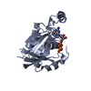


 PDBj
PDBj


 Assembly

 SPODOPTERA FRUGIPERDA (fall armyworm) / References: UniProt: P15153
 Mass: 24.305 Da / Num. of mol.: 1 / Source method: obtained synthetically / Formula: Mg
 Type: RNA linking / Mass: 443.201 Da / Num. of mol.: 1 / Source method: obtained synthetically / Formula: C10H15N5O11P2 / Comment: GDP, energy-carrying molecule*YM
 Mass: 18.015 Da / Num. of mol.: 137 / Source method: isolated from a natural source / Formula: H2O
 Sample preparation
 / Beamline: I02 / Wavelength: 0.9796
  Processing