 Movie
Movie Controller

[English] 日本語
 Yorodumi
- PDB-1gwn: The crystal structure of the core domain of RhoE/Rnd3 - a constit... -
+ Open data

- Basic information
| Entry | Database: PDB / ID: 1gwn | ||||||
|---|---|---|---|---|---|---|---|
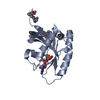
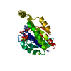
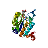
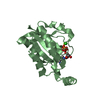
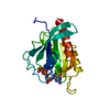
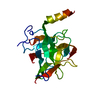
| Title | The crystal structure of the core domain of RhoE/Rnd3 - a constitutively activated small G protein | ||||||
 Components Components | Rho-related GTP-binding protein RhoE | ||||||
 Keywords Keywords | GTPASE / INACTIVE GTPASE / SIGNAL TRANSDUCTION | ||||||
| Function / homology |  Function and homology information Function and homology informationRND3 GTPase cycle / small GTPase-mediated signal transduction / actin filament organization / regulation of actin cytoskeleton organization / Golgi membrane / GTPase activity / protein kinase binding / GTP binding / signal transduction / plasma membrane / cytosol Similarity search - Function | ||||||
| Biological species |  | ||||||
| Method |  X-RAY DIFFRACTION / SYNCHROTRON / MOLECULAR REPLACEMENT / Resolution: 2.1 Å X-RAY DIFFRACTION / SYNCHROTRON / MOLECULAR REPLACEMENT / Resolution: 2.1 Å | ||||||
 Authors Authors | Garavini, H. / Riento, K. / Phelan, J.P. / McAlister, M.S.B. / Ridley, A.J. / Keep, N.H. | ||||||
 Citation Citation | Journal: Biochemistry / Year: 2002 Title: Crystal Structure of the Core Domain of Rhoe/Rnd3: A Constitutively Activated Small G Protein Authors: Garavini, H. / Riento, K. / Phelan, J.P. / McAlister, M.S.B. / Ridley, A.J. / Keep, N.H. | ||||||
| History |
|
- Structure visualization
| Structure viewer | Molecule: MolmilJmol/JSmol |
|---|
- Downloads & links
-Download
| PDBx/mmCIF format | 1gwn.cif.gz | 90.5 KB | Display | PDBx/mmCIF format |
|---|---|---|---|---|
| PDB format | pdb1gwn.ent.gz | 68.6 KB | Display | PDB format |
| PDBx/mmJSON format | 1gwn.json.gz | Tree view | PDBx/mmJSON format | |
| Others |  Other downloads Other downloads |
-Validation report
| Arichive directory | https://data.pdbj.org/pub/pdb/validation_reports/gw/1gwnftp://data.pdbj.org/pub/pdb/validation_reports/gw/1gwn | HTTPS FTP |
|---|
-Related structure data
| Related structure data | 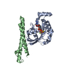 1cxzS S: Starting model for refinement |
|---|---|
| Similar structure data |


-Links
 PDBj
PDBj
- Assembly
| Deposited unit | 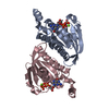
| ||||||||
|---|---|---|---|---|---|---|---|---|---|
| 1 | 
| ||||||||
| 2 | 
| ||||||||
| Unit cell |
| ||||||||
| Noncrystallographic symmetry (NCS) | NCS oper: (Code: given Matrix: (0.978372, -0.026484, 0.205153), Vector: |
-Components
| #1: Protein | Mass: 22862.746 Da / Num. of mol.: 2 / Fragment: CORE DOMAIN, RESIDUES 16-200 Source method: isolated from a genetically manipulated source Source: (gene. exp.)  #2: Chemical |   Mass: 523.180 Da / Num. of mol.: 2 / Source method: obtained synthetically / Formula: C10H16N5O14P3 / Comment: GTP, energy-carrying molecule*YM Mass: 523.180 Da / Num. of mol.: 2 / Source method: obtained synthetically / Formula: C10H16N5O14P3 / Comment: GTP, energy-carrying molecule*YM#3: Chemical |   Mass: 24.305 Da / Num. of mol.: 2 / Source method: obtained synthetically / Formula: Mg Mass: 24.305 Da / Num. of mol.: 2 / Source method: obtained synthetically / Formula: Mg#4: Water | ChemComp-HOH / |  Mass: 18.015 Da / Num. of mol.: 197 / Source method: isolated from a natural source / Formula: H2O Mass: 18.015 Da / Num. of mol.: 197 / Source method: isolated from a natural source / Formula: H2OHas protein modification | Y | Sequence details | RESIDUES 1-15 AND 201 ONWARDS HAVE BEEN DELETED. THE N-TERMINUS INCLUDES AN UNCLEAVED HIS-TAG. | |
|---|
-Experimental details
-Experiment
| Experiment | Method: X-RAY DIFFRACTION / Number of used crystals: 1 |
|---|
- Sample preparation
| Crystal | Density Matthews: 2.25 Å3/Da / Density % sol: 44 % | ||||||||||||||||||||||||||||||
|---|---|---|---|---|---|---|---|---|---|---|---|---|---|---|---|---|---|---|---|---|---|---|---|---|---|---|---|---|---|---|---|
| Crystal grow | pH: 7.5 / Details: 0.1M HEPES PH 7.5, 10% PROPAN-2-OL, 20% PEG 4000 | ||||||||||||||||||||||||||||||
| Crystal grow | *PLUS Temperature: 16 ℃ / Method: vapor diffusion | ||||||||||||||||||||||||||||||
| Components of the solutions | *PLUS
|
-Data collection
| Diffraction | Mean temperature: 100 K |
|---|---|
| Diffraction source | Source: SYNCHROTRON / Site: ESRF  / Beamline: ID14-2 / Wavelength: 0.933 / Beamline: ID14-2 / Wavelength: 0.933 |
| Detector | Type: ADSC CCD / Detector: CCD / Date: Jun 15, 2001 |
| Radiation | Protocol: SINGLE WAVELENGTH / Monochromatic (M) / Laue (L): M / Scattering type: x-ray |
| Radiation wavelength | Wavelength: 0.933 Å / Relative weight: 1 |
| Reflection | Resolution: 2.1→18.8 Å / Num. obs: 25276 / % possible obs: 99.8 % / Redundancy: 4.5 % / Rmerge(I) obs: 0.079 / Net I/σ(I): 6.4 |
| Reflection shell | Resolution: 2.1→2.2 Å / Redundancy: 4.2 % / Rmerge(I) obs: 0.343 / Mean I/σ(I) obs: 2.2 / % possible all: 100 |
| Reflection | *PLUS Num. measured all: 115552 |
| Reflection shell | *PLUS % possible obs: 100 % / Num. unique obs: 3592 / Num. measured obs: 15240 / Mean I/σ(I) obs: 3.5 |
- Processing
| Software |
| ||||||||||||||||||||||||||||||||||||||||||||||||||||||||||||||||||||||||||||||||||||||||||||||||||||||||||||||||||||||||||||||||||||||||||||||||||||||||||||||||||||||||||||||||||||||
|---|---|---|---|---|---|---|---|---|---|---|---|---|---|---|---|---|---|---|---|---|---|---|---|---|---|---|---|---|---|---|---|---|---|---|---|---|---|---|---|---|---|---|---|---|---|---|---|---|---|---|---|---|---|---|---|---|---|---|---|---|---|---|---|---|---|---|---|---|---|---|---|---|---|---|---|---|---|---|---|---|---|---|---|---|---|---|---|---|---|---|---|---|---|---|---|---|---|---|---|---|---|---|---|---|---|---|---|---|---|---|---|---|---|---|---|---|---|---|---|---|---|---|---|---|---|---|---|---|---|---|---|---|---|---|---|---|---|---|---|---|---|---|---|---|---|---|---|---|---|---|---|---|---|---|---|---|---|---|---|---|---|---|---|---|---|---|---|---|---|---|---|---|---|---|---|---|---|---|---|---|---|---|---|
| Refinement | Method to determine structure: MOLECULAR REPLACEMENT Starting model: 1CXZ CHAIN A Resolution: 2.1→18.8 Å / Cor.coef. Fo:Fc: 0.955 / Cor.coef. Fo:Fc free: 0.943 / SU B: 5.871 / SU ML: 0.158 / TLS residual ADP flag: LIKELY RESIDUAL / Cross valid method: THROUGHOUT / ESU R: 0.198 / ESU R Free: 0.161 / Stereochemistry target values: MAXIMUM LIKELIHOOD Details: HYDROGENS HAVE BEEN ADDED IN THE RIDING POSITIONS. THE ELECTRON DENSITY IN THE REGION OF RESIDUE 23 IS AMBIGUOUS AND THE CIS-CONFORMATION OF LYS23 MAY BE DUE TO THE POOR DEFINITION OF THE ENCLOSING DENSITY.
| ||||||||||||||||||||||||||||||||||||||||||||||||||||||||||||||||||||||||||||||||||||||||||||||||||||||||||||||||||||||||||||||||||||||||||||||||||||||||||||||||||||||||||||||||||||||
| Solvent computation | Ion probe radii: 0.8 Å / Shrinkage radii: 0.8 Å / VDW probe radii: 1.4 Å / Solvent model: BABINET MODEL WITH MASK | ||||||||||||||||||||||||||||||||||||||||||||||||||||||||||||||||||||||||||||||||||||||||||||||||||||||||||||||||||||||||||||||||||||||||||||||||||||||||||||||||||||||||||||||||||||||
| Refinement step | Cycle: LAST / Resolution: 2.1→18.8 Å
| ||||||||||||||||||||||||||||||||||||||||||||||||||||||||||||||||||||||||||||||||||||||||||||||||||||||||||||||||||||||||||||||||||||||||||||||||||||||||||||||||||||||||||||||||||||||
| Refine LS restraints |
|