regulation of mast cell chemotaxis / regulation of cell-substrate adhesion / lymphocyte aggregation / positive regulation of mast cell proliferation / regulation of respiratory burst / regulation of mast cell degranulation / mast cell proliferation / erythrocyte enucleation / regulation of neutrophil migration / NADPH oxidase complex ...regulation of mast cell chemotaxis / regulation of cell-substrate adhesion / lymphocyte aggregation / positive regulation of mast cell proliferation / regulation of respiratory burst / regulation of mast cell degranulation / mast cell proliferation / erythrocyte enucleation / regulation of neutrophil migration / NADPH oxidase complex / regulation of hydrogen peroxide metabolic process / respiratory burst / cortical cytoskeleton organization / cell projection assembly / protein kinase regulator activity / ROS and RNS production in phagocytes / PCP/CE pathway / positive regulation of neutrophil chemotaxis / superoxide anion generation / : / small GTPase-mediated signal transduction / regulation of T cell proliferation / establishment or maintenance of cell polarity / RAC2 GTPase cycle / RHO GTPases Activate NADPH Oxidases / bone resorption / GPVI-mediated activation cascade / positive regulation of lamellipodium assembly / cell projection / actin filament organization / small monomeric GTPase / regulation of actin cytoskeleton organization / phagocytic vesicle membrane / chemotaxis / Constitutive Signaling by Aberrant PI3K in Cancer / PIP3 activates AKT signaling / nuclear envelope / regulation of cell shape / lamellipodium / PI5P, PP2A and IER3 Regulate PI3K/AKT Signaling / G protein activity / cytoplasmic vesicle / cytoskeleton / mitochondrial outer membrane / G protein-coupled receptor signaling pathway / focal adhesion / GTPase activity / protein kinase binding / endoplasmic reticulum membrane / GTP binding / signal transduction / extracellular exosome / plasma membrane / cytosol Similarity search - Function
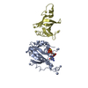
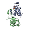
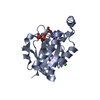
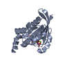
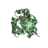
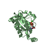
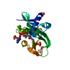
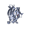
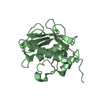
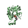
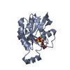
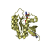
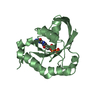
Small GTPase Rho / Small GTPase Rho domain profile. / Ran (Ras-related nuclear proteins) /TC4 subfamily of small GTPases / Rho (Ras homology) subfamily of Ras-like small GTPases / Ras subfamily of RAS small GTPases / Small GTPase / Ras family / Rab subfamily of small GTPases / Small GTP-binding protein domain / P-loop containing nucleotide triphosphate hydrolases ...Small GTPase Rho / Small GTPase Rho domain profile. / Ran (Ras-related nuclear proteins) /TC4 subfamily of small GTPases / Rho (Ras homology) subfamily of Ras-like small GTPases / Ras subfamily of RAS small GTPases / Small GTPase / Ras family / Rab subfamily of small GTPases / Small GTP-binding protein domain / P-loop containing nucleotide triphosphate hydrolases / Rossmann fold / P-loop containing nucleoside triphosphate hydrolase / 3-Layer(aba) Sandwich / Alpha Beta Similarity search - Domain/homology
Mass: 18.015 Da / Num. of mol.: 527 / Source method: isolated from a natural source / Formula: H2O
Compound details
ENGINEERED RESIDUE IN CHAIN A, GLY 12 TO VAL ENGINEERED RESIDUE IN CHAIN B, GLY 12 TO VAL ...ENGINEERED RESIDUE IN CHAIN A, GLY 12 TO VAL ENGINEERED RESIDUE IN CHAIN B, GLY 12 TO VAL ENGINEERED RESIDUE IN CHAIN C, GLY 12 TO VAL ENGINEERED RESIDUE IN CHAIN D, GLY 12 TO VAL
Sequence details
THERE IS TAG LINKER SEQUENCE, GGGSGGS (NOT SEEN) AND A MUTATION G12V
-
Experimental details
-
Experiment
Experiment
Method: X-RAY DIFFRACTION / Number of used crystals: 1
-
Sample preparation
Crystal
Density Matthews: 2.7 Å3/Da / Density % sol: 54 % / Description: NONE
Crystal grow
Temperature: 293 K / pH: 7 Details: RAC2(2-177)GTPGS WAS CRYSTALLIZED USING A PROTEIN CONCENTRATION OF 25 MG/ML WITH PRECIPITANT (18% PEG3350, 300 MM SODIUM/POTASSIUM TARTRATE) AND AT A CONSTANT TEMPERATURE OF 20C
Protocol: SINGLE WAVELENGTH / Monochromatic (M) / Laue (L): M / Scattering type: x-ray
Radiation wavelength
Wavelength: 0.9796 Å / Relative weight: 1
Reflection
Resolution: 1.95→54 Å / Num. obs: 79004 / % possible obs: 98.7 % / Redundancy: 4.8 % / Biso Wilson estimate: 20.98 Å2 / Rmerge(I) obs: 0.08 / Net I/σ(I): 12.9
Reflection shell
Resolution: 1.95→2.06 Å / Redundancy: 4.5 % / Rmerge(I) obs: 0.52 / Mean I/σ(I) obs: 2.3 / % possible all: 96.1
-
Processing
Software
Name
Version
Classification
PHENIX
(PHENIX.REFINE)
refinement
MOSFLM
datareduction
SCALA
datascaling
PHASER
phasing
Refinement
Method to determine structure: MOLECULAR REPLACEMENT Starting model: IN HOUSE RAC2 STRUCTURE Resolution: 2→53.62 Å / SU ML: 0.45 / σ(F): 1.12 / Phase error: 40.6 / Stereochemistry target values: ML Details: DATA INITIALLY THOUGHT TO BE P212121, LATER FOUND TO BE P21 WITH B AS 90, ALSO TWINNED.
Rfactor
Num. reflection
% reflection
Rfree
0.345
4720
5.1 %
Rwork
0.28
-
-
obs
0.283
92859
82.9 %
Solvent computation
Shrinkage radii: 0.9 Å / VDW probe radii: 1.11 Å / Solvent model: FLAT BULK SOLVENT MODEL / Bsol: 50.57 Å2 / ksol: 0.35 e/Å3
Displacement parameters
Baniso -1
Baniso -2
Baniso -3
1-
-8.9782 Å2
0 Å2
0.7393 Å2
2-
-
23.0459 Å2
0 Å2
3-
-
-
-14.0677 Å2
Refinement step
Cycle: LAST / Resolution: 2→53.62 Å
Protein
Nucleic acid
Ligand
Solvent
Total
Num. atoms
5156
0
128
527
5811
Refine LS restraints
Refine-ID
Type
Dev ideal
Number
X-RAY DIFFRACTION
f_bond_d
0.002
5442
X-RAY DIFFRACTION
f_angle_d
0.641
7478
X-RAY DIFFRACTION
f_dihedral_angle_d
18.544
1907
X-RAY DIFFRACTION
f_chiral_restr
0.042
894
X-RAY DIFFRACTION
f_plane_restr
0.003
921
LS refinement shell
Resolution (Å)
Rfactor Rfree
Num. reflection Rfree
Rfactor Rwork
Num. reflection Rwork
Refine-ID
% reflection obs (%)
2-2.0227
0.4149
138
0.3968
2645
X-RAY DIFFRACTION
74
2.0227-2.0465
0.4833
141
0.4161
2751
X-RAY DIFFRACTION
79
2.0465-2.0715
0.4714
162
0.3996
2794
X-RAY DIFFRACTION
79
2.0715-2.0977
0.4484
162
0.3819
2819
X-RAY DIFFRACTION
78
2.0977-2.1253
0.4401
157
0.3718
2757
X-RAY DIFFRACTION
79
2.1253-2.1544
0.4093
135
0.3619
2771
X-RAY DIFFRACTION
79
2.1544-2.1852
0.3706
142
0.3384
2837
X-RAY DIFFRACTION
79
2.1852-2.2178
0.4115
156
0.3282
2837
X-RAY DIFFRACTION
80
2.2178-2.2525
0.4475
153
0.3602
2752
X-RAY DIFFRACTION
78
2.2525-2.2894
0.375
125
0.3244
2804
X-RAY DIFFRACTION
80
2.2894-2.3289
0.3522
155
0.3177
2943
X-RAY DIFFRACTION
81
2.3289-2.3712
0.4049
179
0.3037
2766
X-RAY DIFFRACTION
80
2.3712-2.4169
0.3734
155
0.2941
2843
X-RAY DIFFRACTION
81
2.4169-2.4662
0.3081
163
0.2837
2939
X-RAY DIFFRACTION
82
2.4662-2.5198
0.3707
150
0.2884
2884
X-RAY DIFFRACTION
81
2.5198-2.5784
0.3563
165
0.3086
2901
X-RAY DIFFRACTION
83
2.5784-2.6429
0.4088
121
0.3307
3066
X-RAY DIFFRACTION
84
2.6429-2.7144
0.3911
163
0.2986
2898
X-RAY DIFFRACTION
84
2.7144-2.7942
0.3631
139
0.3059
3046
X-RAY DIFFRACTION
85
2.7942-2.8844
0.3906
148
0.3019
3036
X-RAY DIFFRACTION
86
2.8844-2.9875
0.408
182
0.2943
3049
X-RAY DIFFRACTION
86
2.9875-3.1071
0.364
158
0.2762
3066
X-RAY DIFFRACTION
87
3.1071-3.2485
0.3488
180
0.253
3181
X-RAY DIFFRACTION
88
3.2485-3.4197
0.2958
180
0.2402
2988
X-RAY DIFFRACTION
87
3.4197-3.6339
0.2808
179
0.2418
3103
X-RAY DIFFRACTION
87
3.6339-3.9144
0.3147
146
0.2314
3144
X-RAY DIFFRACTION
89
3.9144-4.3082
0.2524
166
0.2286
3147
X-RAY DIFFRACTION
88
4.3082-4.9313
0.2707
205
0.1991
3092
X-RAY DIFFRACTION
88
4.9313-6.2114
0.2786
163
0.2313
3162
X-RAY DIFFRACTION
89
6.2114-53.6375
0.2898
152
0.2259
3118
X-RAY DIFFRACTION
87
+
About Yorodumi
-
News
-
Feb 9, 2022. New format data for meta-information of EMDB entries
New format data for meta-information of EMDB entries
Version 3 of the EMDB header file is now the official format.
The previous official version 1.9 will be removed from the archive.
In the structure databanks used in Yorodumi, some data are registered as the other names, "COVID-19 virus" and "2019-nCoV". Here are the details of the virus and the list of structure data.
Jan 31, 2019. EMDB accession codes are about to change! (news from PDBe EMDB page)
EMDB accession codes are about to change! (news from PDBe EMDB page)
The allocation of 4 digits for EMDB accession codes will soon come to an end. Whilst these codes will remain in use, new EMDB accession codes will include an additional digit and will expand incrementally as the available range of codes is exhausted. The current 4-digit format prefixed with “EMD-” (i.e. EMD-XXXX) will advance to a 5-digit format (i.e. EMD-XXXXX), and so on. It is currently estimated that the 4-digit codes will be depleted around Spring 2019, at which point the 5-digit format will come into force.
The EM Navigator/Yorodumi systems omit the EMD- prefix.
Related info.:Q: What is EMD? / ID/Accession-code notation in Yorodumi/EM Navigator
Yorodumi is a browser for structure data from EMDB, PDB, SASBDB, etc.
This page is also the successor to EM Navigator detail page, and also detail information page/front-end page for Omokage search.
The word "yorodu" (or yorozu) is an old Japanese word meaning "ten thousand". "mi" (miru) is to see.
Related info.:EMDB / PDB / SASBDB / Comparison of 3 databanks / Yorodumi Search / Aug 31, 2016. New EM Navigator & Yorodumi / Yorodumi Papers / Jmol/JSmol / Function and homology information / Changes in new EM Navigator and Yorodumi
 Movie
Movie Controller


 Open data
 Basic information
 Components
 Keywords
 Function and homology information
 HOMO SAPIENS (human)
 X-RAY DIFFRACTION /
  Authors
 Citation
 Structure visualization
 Downloads & links
 Other downloads
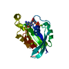

 PDBj
PDBj


 Assembly
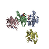
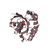
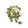

 SPODOPTERA FRUGIPERDA (fall armyworm) / References: UniProt: P15153
 Mass: 523.180 Da / Num. of mol.: 4 / Source method: obtained synthetically / Formula: C10H16N5O14P3 / Comment: GTP, energy-carrying molecule*YM
 Mass: 18.015 Da / Num. of mol.: 527 / Source method: isolated from a natural source / Formula: H2O
 Sample preparation
 / Beamline: I02 / Wavelength: 0.9796
  Processing