 Movie
Movie Controller


+ Open data

- Basic information
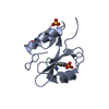
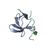
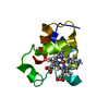
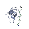
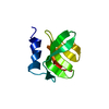
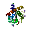
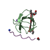
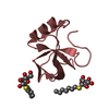
| Entry | Database: PDB / ID: 2w10 | ||||||
|---|---|---|---|---|---|---|---|
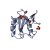
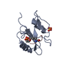
| Title | Mona SH3C in complex | ||||||
 Components Components |
| ||||||
 Keywords Keywords | HYDROLASE / ALTERNATIVE SPLICING / TPR REPEAT / SH2 DOMAIN / SH3 DOMAIN / COILED COIL / PROTEIN PHOSPHATASE / CYTOPLASMIC VESICLE / PHOSPHOPROTEIN / SIGNAL TRANDUCTION / SH3 DOMAIN-COMPLEX / SH3 / GADS / MONA / DIMER / HD-PTP / CYTOPLASM | ||||||
| Function / homology |  Function and homology information Function and homology informationpositive regulation of adherens junction organization / positive regulation of homophilic cell adhesion / FLT3 Signaling / Co-stimulation by CD28 / FCERI mediated MAPK activation / Signaling by SCF-KIT / Generation of second messenger molecules / positive regulation of Wnt protein secretion / positive regulation of early endosome to late endosome transport / FCERI mediated Ca+2 mobilization ...positive regulation of adherens junction organization / positive regulation of homophilic cell adhesion / FLT3 Signaling / Co-stimulation by CD28 / FCERI mediated MAPK activation / Signaling by SCF-KIT / Generation of second messenger molecules / positive regulation of Wnt protein secretion / positive regulation of early endosome to late endosome transport / FCERI mediated Ca+2 mobilization / negative regulation of epithelial cell migration / protein transport to vacuole involved in ubiquitin-dependent protein catabolic process via the multivesicular body sorting pathway / early endosome to late endosome transport / endocytic recycling / ubiquitin-dependent protein catabolic process via the multivesicular body sorting pathway / DAP12 signaling / regulation of MAPK cascade / cilium assembly / phosphotyrosine residue binding / protein-tyrosine-phosphatase / protein tyrosine phosphatase activity / centriolar satellite / early endosome / endosome / nuclear body / ciliary basal body / protein kinase binding / signal transduction / nucleoplasm / nucleus / plasma membrane / cytoplasm / cytosol Similarity search - Function | ||||||
| Biological species |  | ||||||
| Method |  X-RAY DIFFRACTION / SYNCHROTRON / MOLECULAR REPLACEMENT / Resolution: 1.9 Å X-RAY DIFFRACTION / SYNCHROTRON / MOLECULAR REPLACEMENT / Resolution: 1.9 Å | ||||||
 Authors Authors | Harkiolaki, M. / Feller, S.M. | ||||||
 Citation Citation | Journal: Structure / Year: 2009 Title: Distinct Binding Modes of Two Epitopes in Gab2 that Interact with the Sh3C Domain of Grb2. Authors: Harkiolaki, M. / Tsirka, T. / Lewitzky, M. / Simister, P.C. / Joshi, D. / Bird, L.E. / Jones, E.Y. / O'Reilly, N. / Feller, S.M. | ||||||
| History |
|
- Structure visualization
| Structure viewer | Molecule: MolmilJmol/JSmol |
|---|
- Downloads & links
-Download
| PDBx/mmCIF format | 2w10.cif.gz | 44.3 KB | Display | PDBx/mmCIF format |
|---|---|---|---|---|
| PDB format | pdb2w10.ent.gz | 31.4 KB | Display | PDB format |
| PDBx/mmJSON format | 2w10.json.gz | Tree view | PDBx/mmJSON format | |
| Others |  Other downloads Other downloads |
-Validation report
| Arichive directory | https://data.pdbj.org/pub/pdb/validation_reports/w1/2w10ftp://data.pdbj.org/pub/pdb/validation_reports/w1/2w10 | HTTPS FTP |
|---|
-Related structure data
| Related structure data | 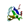 2vvkC  2vwfC 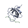 2w0zC  1utiS S: Starting model for refinement C: citing same article ( |
|---|---|
| Similar structure data |


-Links
 PDBj
PDBj


- Assembly
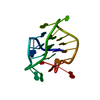
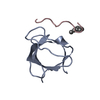
| Deposited unit | 
| ||||||||
|---|---|---|---|---|---|---|---|---|---|
| 1 | 
| ||||||||
| 2 | 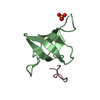
| ||||||||
| Unit cell |
|
-Components
| #1: Protein | Mass: 7088.970 Da / Num. of mol.: 2 / Fragment: SH3 2, RESIDUES 265-322 Source method: isolated from a genetically manipulated source Source: (gene. exp.)  #2: Protein/peptide | Mass: 1285.575 Da / Num. of mol.: 2 / Fragment: SH3 BINDING REGION, RESIDUES 719-730 / Source method: obtained synthetically / Source: (synth.) #3: Chemical |   Mass: 94.971 Da / Num. of mol.: 2 / Source method: obtained synthetically / Formula: PO4 Mass: 94.971 Da / Num. of mol.: 2 / Source method: obtained synthetically / Formula: PO4#4: Water | ChemComp-HOH / |  Mass: 18.015 Da / Num. of mol.: 135 / Source method: isolated from a natural source / Formula: H2O Mass: 18.015 Da / Num. of mol.: 135 / Source method: isolated from a natural source / Formula: H2OSequence details | PLGS OVERHANG DUE TO EXPRESSION | |
|---|
-Experimental details
-Experiment
| Experiment | Method: X-RAY DIFFRACTION / Number of used crystals: 1 |
|---|
- Sample preparation
| Crystal | Density Matthews: 1.67 Å3/Da / Density % sol: 25.96 % / Description: NONE |
|---|---|
| Crystal grow | pH: 7.5 Details: 0.1 M PHOSPHATE CITRATE, 2 M AMMONIUM SULPHATE, pH 7.5 |
-Data collection
| Diffraction | Mean temperature: 77 K |
|---|---|
| Diffraction source | Source: SYNCHROTRON / Site: ESRF  / Beamline: BM14 / Wavelength: 0.978 / Beamline: BM14 / Wavelength: 0.978 |
| Detector | Type: MARRESEARCH / Detector: CCD / Date: Apr 15, 2006 / Details: MIRRORS |
| Radiation | Monochromator: SI(111) / Protocol: SINGLE WAVELENGTH / Monochromatic (M) / Laue (L): M / Scattering type: x-ray |
| Radiation wavelength | Wavelength: 0.978 Å / Relative weight: 1 |
| Reflection | Resolution: 1.86→30 Å / Num. obs: 8966 / % possible obs: 81.9 % / Observed criterion σ(I): 1 / Redundancy: 2 % / Rmerge(I) obs: 0.04 / Net I/σ(I): 18.7 |
| Reflection shell | Resolution: 1.86→1.94 Å / Redundancy: 2 % / Rmerge(I) obs: 0.06 / Mean I/σ(I) obs: 11.5 / % possible all: 44.2 |
- Processing
| Software |
| ||||||||||||||||||||||||||||||||||||||||||||||||||||||||||||||||||||||||||||||||||||||||||||||||||||||||||||||||||||||||||||||||||||||||||||||||||||||||||||||||||||||||||||||||||||||
|---|---|---|---|---|---|---|---|---|---|---|---|---|---|---|---|---|---|---|---|---|---|---|---|---|---|---|---|---|---|---|---|---|---|---|---|---|---|---|---|---|---|---|---|---|---|---|---|---|---|---|---|---|---|---|---|---|---|---|---|---|---|---|---|---|---|---|---|---|---|---|---|---|---|---|---|---|---|---|---|---|---|---|---|---|---|---|---|---|---|---|---|---|---|---|---|---|---|---|---|---|---|---|---|---|---|---|---|---|---|---|---|---|---|---|---|---|---|---|---|---|---|---|---|---|---|---|---|---|---|---|---|---|---|---|---|---|---|---|---|---|---|---|---|---|---|---|---|---|---|---|---|---|---|---|---|---|---|---|---|---|---|---|---|---|---|---|---|---|---|---|---|---|---|---|---|---|---|---|---|---|---|---|---|
| Refinement | Method to determine structure: MOLECULAR REPLACEMENT Starting model: PDB ENTRY 1UTI Resolution: 1.9→35.78 Å / Cor.coef. Fo:Fc: 0.956 / Cor.coef. Fo:Fc free: 0.887 / SU B: 3.927 / SU ML: 0.117 / Cross valid method: THROUGHOUT / ESU R: 0.197 / ESU R Free: 0.177 / Stereochemistry target values: MAXIMUM LIKELIHOOD Details: HYDROGENS HAVE BEEN ADDED IN THE RIDING POSITIONS. U VALUES REFINED INDIVIDUALLY.
| ||||||||||||||||||||||||||||||||||||||||||||||||||||||||||||||||||||||||||||||||||||||||||||||||||||||||||||||||||||||||||||||||||||||||||||||||||||||||||||||||||||||||||||||||||||||
| Solvent computation | Ion probe radii: 0.8 Å / Shrinkage radii: 0.8 Å / VDW probe radii: 1.4 Å / Solvent model: MASK | ||||||||||||||||||||||||||||||||||||||||||||||||||||||||||||||||||||||||||||||||||||||||||||||||||||||||||||||||||||||||||||||||||||||||||||||||||||||||||||||||||||||||||||||||||||||
| Displacement parameters | Biso mean: 12.28 Å2
| ||||||||||||||||||||||||||||||||||||||||||||||||||||||||||||||||||||||||||||||||||||||||||||||||||||||||||||||||||||||||||||||||||||||||||||||||||||||||||||||||||||||||||||||||||||||
| Refinement step | Cycle: LAST / Resolution: 1.9→35.78 Å
| ||||||||||||||||||||||||||||||||||||||||||||||||||||||||||||||||||||||||||||||||||||||||||||||||||||||||||||||||||||||||||||||||||||||||||||||||||||||||||||||||||||||||||||||||||||||
| Refine LS restraints |
|