 Movie
Movie Controller

[English] 日本語
 Yorodumi
- PDB-2d0n: Crystal structure of the C-terminal SH3 domain of the adaptor pro... -
+ Open data

- Basic information
| Entry | Database: PDB / ID: 2d0n | ||||||
|---|---|---|---|---|---|---|---|
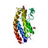
| Title | Crystal structure of the C-terminal SH3 domain of the adaptor protein GADS in complex with SLP-76 motif peptide reveals a unique SH3-SH3 interaction | ||||||
 Components Components |
| ||||||
 Keywords Keywords | SIGNALING PROTEIN / SH3 DOMAIN-COMPLEX / MONA/GADS SH3C DOMAIN | ||||||
| Function / homology |  Function and homology information Function and homology informationFLT3 Signaling / Co-stimulation by CD28 / FCERI mediated MAPK activation / Signaling by SCF-KIT / Generation of second messenger molecules / FCERI mediated Ca+2 mobilization / DAP12 signaling / regulation of MAPK cascade / phosphotyrosine residue binding / endosome ...FLT3 Signaling / Co-stimulation by CD28 / FCERI mediated MAPK activation / Signaling by SCF-KIT / Generation of second messenger molecules / FCERI mediated Ca+2 mobilization / DAP12 signaling / regulation of MAPK cascade / phosphotyrosine residue binding / endosome / signal transduction / nucleoplasm / nucleus / plasma membrane / cytoplasm / cytosol Similarity search - Function | ||||||
| Biological species |  | ||||||
| Method |  X-RAY DIFFRACTION / SYNCHROTRON / MOLECULAR REPLACEMENT / Resolution: 1.57 Å X-RAY DIFFRACTION / SYNCHROTRON / MOLECULAR REPLACEMENT / Resolution: 1.57 Å | ||||||
 Authors Authors | Dimasi, N. | ||||||
 Citation Citation | Journal: Int.J.Biochem.Cell Biol. / Year: 2007 Title: Crystal structure of the C-terminal SH3 domain of the adaptor protein GADS in complex with SLP-76 motif peptide reveals a unique SH3-SH3 interaction Authors: Dimasi, N. | ||||||
| History |
|
- Structure visualization
| Structure viewer | Molecule: MolmilJmol/JSmol |
|---|
- Downloads & links
-Download
| PDBx/mmCIF format | 2d0n.cif.gz | 43.7 KB | Display | PDBx/mmCIF format |
|---|---|---|---|---|
| PDB format | pdb2d0n.ent.gz | 30.5 KB | Display | PDB format |
| PDBx/mmJSON format | 2d0n.json.gz | Tree view | PDBx/mmJSON format | |
| Others |  Other downloads Other downloads |
-Validation report
| Arichive directory | https://data.pdbj.org/pub/pdb/validation_reports/d0/2d0nftp://data.pdbj.org/pub/pdb/validation_reports/d0/2d0n | HTTPS FTP |
|---|
-Related structure data
| Related structure data |  1oebS S: Starting model for refinement |
|---|---|
| Similar structure data |


-Links
 PDBj
PDBj


- Assembly
| Deposited unit | 
| ||||||||||||||||||
|---|---|---|---|---|---|---|---|---|---|---|---|---|---|---|---|---|---|---|---|
| 1 |
| ||||||||||||||||||
| Unit cell |
| ||||||||||||||||||
| Components on special symmetry positions |
|
-Components
| #1: Protein | Mass: 6719.488 Da / Num. of mol.: 2 / Fragment: C-terminal SH3 domain Source method: isolated from a genetically manipulated source Source: (gene. exp.)  #2: Protein/peptide | Mass: 1002.123 Da / Num. of mol.: 2 / Source method: obtained synthetically Details: The SLP-76 peptide was synthesized synthetically with the F-moc chemistry #3: Water | ChemComp-HOH / |  Mass: 18.015 Da / Num. of mol.: 199 / Source method: isolated from a natural source / Formula: H2O Mass: 18.015 Da / Num. of mol.: 199 / Source method: isolated from a natural source / Formula: H2O |
|---|
-Experimental details
-Experiment
| Experiment | Method: X-RAY DIFFRACTION / Number of used crystals: 1 |
|---|
- Sample preparation
| Crystal | Density Matthews: 2.08 Å3/Da / Density % sol: 42.04 % |
|---|---|
| Crystal grow | Temperature: 298 K / Method: vapor diffusion, hanging drop / pH: 6.5 Details: 1.26M ammonium sulfate, 0.1M cacodylate, pH 6.5, VAPOR DIFFUSION, HANGING DROP, temperature 298.0K |
-Data collection
| Diffraction | Mean temperature: 100 K |
|---|---|
| Diffraction source | Source: SYNCHROTRON / Site: NSLS  / Beamline: X8C / Wavelength: 0.9 Å / Beamline: X8C / Wavelength: 0.9 Å |
| Detector | Type: ADSC QUANTUM 4 / Detector: CCD / Date: Apr 13, 2005 |
| Radiation | Monochromator: MIRRORS / Protocol: SINGLE WAVELENGTH / Monochromatic (M) / Laue (L): M / Scattering type: x-ray |
| Radiation wavelength | Wavelength: 0.9 Å / Relative weight: 1 |
| Reflection | Resolution: 1.54→36.18 Å / Num. obs: 16893 / % possible obs: 85.1 % / Observed criterion σ(F): 2 / Observed criterion σ(I): 2 / Rmerge(I) obs: 0.043 / Rsym value: 0.178 |
| Reflection shell | Resolution: 1.54→1.59 Å / Rmerge(I) obs: 0.043 / % possible all: 85.1 |
- Processing
| Software |
| ||||||||||||||||||||
|---|---|---|---|---|---|---|---|---|---|---|---|---|---|---|---|---|---|---|---|---|---|
| Refinement | Method to determine structure: MOLECULAR REPLACEMENT Starting model: PDB ENTRY 1OEB Chain A Resolution: 1.57→7.9 Å / σ(F): 2 / Stereochemistry target values: Engh & Huber
| ||||||||||||||||||||
| Refinement step | Cycle: LAST / Resolution: 1.57→7.9 Å
| ||||||||||||||||||||
| LS refinement shell | Highest resolution: 1.57 Å |
