[English] 日本語
Yorodumi
- PDB-3c5r: Crystal Structure of the BARD1 Ankyrin Repeat Domain and Its Func... -

+
Open data


ID or keywords:

Loading...

-
Basic information

Entry
Database: PDB / ID: 3c5r
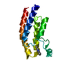
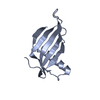
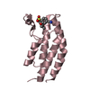
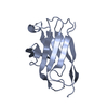
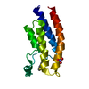
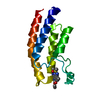
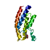
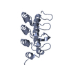
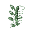
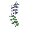
TitleCrystal Structure of the BARD1 Ankyrin Repeat Domain and Its Functional Consequences
ComponentsBRCA1-associated RING domain protein 1
KeywordsPROTEIN BINDING / BARD1 / Ankyrin repeat / helix / extended loop / four repeat / protein / ANK repeat / Disease mutation / Metal-binding / Nucleus / Zinc-finger
Function / homology
Function and homology information


negative regulation of mRNA 3'-end processing / Defective DNA double strand break response due to BRCA1 loss of function / Defective DNA double strand break response due to BARD1 loss of function / BRCA1-BARD1 complex / BRCA1-C complex / BRCA1-B complex / BRCA1-A complex / nuclear ubiquitin ligase complex / DNA strand resection involved in replication fork processing / homologous recombination ...negative regulation of mRNA 3'-end processing / Defective DNA double strand break response due to BRCA1 loss of function / Defective DNA double strand break response due to BARD1 loss of function / BRCA1-BARD1 complex / BRCA1-C complex / BRCA1-B complex / BRCA1-A complex / nuclear ubiquitin ligase complex / DNA strand resection involved in replication fork processing / homologous recombination / tissue homeostasis / protein K6-linked ubiquitination / regulation of DNA damage checkpoint / regulation of phosphorylation / Impaired BRCA2 binding to PALB2 / mitotic G2/M transition checkpoint / negative regulation of protein export from nucleus / Defective homologous recombination repair (HRR) due to BRCA1 loss of function / Defective HDR through Homologous Recombination Repair (HRR) due to PALB2 loss of BRCA1 binding function / Defective HDR through Homologous Recombination Repair (HRR) due to PALB2 loss of BRCA2/RAD51/RAD51C binding function / Homologous DNA Pairing and Strand Exchange / Resolution of D-loop Structures through Synthesis-Dependent Strand Annealing (SDSA) / Resolution of D-loop Structures through Holliday Junction Intermediates / HDR through Single Strand Annealing (SSA) / Impaired BRCA2 binding to RAD51 / Presynaptic phase of homologous DNA pairing and strand exchange / negative regulation of cell cycle / regulation of DNA repair / ubiquitin ligase complex / cellular response to ionizing radiation / Nonhomologous End-Joining (NHEJ) / RING-type E3 ubiquitin transferase / HDR through Homologous Recombination (HRR) / G2/M DNA damage checkpoint / Metalloprotease DUBs / kinase binding / cytoplasmic ribonucleoprotein granule / positive regulation of protein catabolic process / ubiquitin-protein transferase activity / UCH proteinases / Recruitment and ATM-mediated phosphorylation of repair and signaling proteins at DNA double strand breaks / Processing of DNA double-strand break ends / Regulation of TP53 Activity through Phosphorylation / regulation of cell cycle / protein ubiquitination / nuclear speck / positive regulation of apoptotic process / protein heterodimerization activity / DNA repair / DNA damage response / negative regulation of apoptotic process / protein homodimerization activity / RNA binding / nucleoplasm / nucleus / metal ion binding / cytoplasm
Similarity search - Function
BARD1, Zinc finger, RING-type / zf-RING of BARD1-type protein / Ankyrin repeat-containing domain / BRCA1 C Terminus (BRCT) domain / breast cancer carboxy-terminal domain / Zinc finger, RING-type, conserved site / Zinc finger RING-type signature. / BRCT domain profile. / BRCT domain / BRCT domain superfamily ...BARD1, Zinc finger, RING-type / zf-RING of BARD1-type protein / Ankyrin repeat-containing domain / BRCA1 C Terminus (BRCT) domain / breast cancer carboxy-terminal domain / Zinc finger, RING-type, conserved site / Zinc finger RING-type signature. / BRCT domain profile. / BRCT domain / BRCT domain superfamily / Ankyrin repeat / Ankyrin repeats (3 copies) / Ankyrin repeat profile. / Ankyrin repeat region circular profile. / ankyrin repeats / Ankyrin repeat / Zinc finger RING-type profile. / Zinc finger, RING-type / Ankyrin repeat-containing domain superfamily / Serine Threonine Protein Phosphatase 5, Tetratricopeptide repeat / Alpha Horseshoe / Zinc finger, RING/FYVE/PHD-type / Mainly Alpha
Similarity search - Domain/homology
BRCA1-associated RING domain protein 1
Similarity search - Component
Biological speciesHomo sapiens (human)
MethodX-RAY DIFFRACTION / MOLECULAR REPLACEMENT / Resolution: 2 Å
AuthorsFox III, D. / Le Trong, I. / Stenkamp, R.E. / Klevit, R.E.
CitationJournal: J.Biol.Chem. / Year: 2008
Title: Crystal Structure of the BARD1 Ankyrin Repeat Domain and Its Functional Consequences.
Authors: Fox, D. / Le Trong, I. / Rajagopal, P. / Brzovic, P.S. / Stenkamp, R.E. / Klevit, R.E.
History
DepositionFeb 1, 2008Deposition site: RCSB / Processing site: RCSB
Revision 1.0May 13, 2008Provider: repository / Type: Initial release
Revision 1.1Jul 13, 2011Group: Advisory / Version format compliance
Revision 1.2Aug 30, 2023Group: Data collection / Database references / Refinement description
Category: chem_comp_atom / chem_comp_bond ...chem_comp_atom / chem_comp_bond / database_2 / pdbx_initial_refinement_model / struct_ref_seq_dif
Item: _database_2.pdbx_DOI / _database_2.pdbx_database_accession / _struct_ref_seq_dif.details

-
Structure visualization

Structure viewerMolecule:
MolmilJmol/JSmol

Downloads & links

-
Assembly

Deposited unit
A: BRCA1-associated RING domain protein 1
B: BRCA1-associated RING domain protein 1


Theoretical massNumber of molelcules
Total (without water)29,9532
Polymers29,9532
Non-polymers00
Water4,125229
1
A: BRCA1-associated RING domain protein 1


Theoretical massNumber of molelcules
Total (without water)14,9771
Polymers14,9771
Non-polymers00
Water181
TypeNameSymmetry operationNumber
identity operation1_555x,y,z1
2
B: BRCA1-associated RING domain protein 1


Theoretical massNumber of molelcules
Total (without water)14,9771
Polymers14,9771
Non-polymers00
Water181
TypeNameSymmetry operationNumber
identity operation1_555x,y,z1
3
A: BRCA1-associated RING domain protein 1

B: BRCA1-associated RING domain protein 1


Theoretical massNumber of molelcules
Total (without water)29,9532
Polymers29,9532
Non-polymers00
Water362
TypeNameSymmetry operationNumber
identity operation1_555x,y,z1
crystal symmetry operation2_656-x+1,y+1/2,-z+11
Buried area1370 Å2
ΔGint-12 kcal/mol
Surface area11700 Å2
MethodPISA
Unit cell
Length a, b, c (Å)37.847, 74.112, 51.174
Angle α, β, γ (deg.)90.00, 105.23, 90.00
Int Tables number4
Space group name H-MP1211
DetailsThere are 2 biological units in the asymmetric unit (chains A & B)

-
Components

#1: Protein BRCA1-associated RING domain protein 1 / BARD-1


Mass: 14976.739 Da / Num. of mol.: 2
Fragment: BARD1 Ankyrin Repeat Domain (UNP residues 425-555)
Source method: isolated from a genetically manipulated source
Source: (gene. exp.) Homo sapiens (human) / Gene: BARD1 / Plasmid: pET151D / Species (production host): Escherichia coli / Production host: Escherichia coli BL21(DE3) (bacteria) / Strain (production host): BL21(DE3) / References: UniProt: Q99728
#2: Water ChemComp-HOH / water


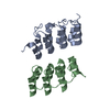
Mass: 18.015 Da / Num. of mol.: 229 / Source method: isolated from a natural source / Formula: H2O

-
Experimental details

-
Experiment

ExperimentMethod: X-RAY DIFFRACTION / Number of used crystals: 1

-
Sample preparation

CrystalDensity Matthews: 2.31 Å3/Da / Density % sol: 46.8 %
Crystal growTemperature: 277 K / Method: vapor diffusion, hanging drop / pH: 8.5
Details: 0.2 Mg Chloride, 0.1 M Tris Hydrochloride pH 8.5, 25% w/v PEG 3350, VAPOR DIFFUSION, HANGING DROP, temperature 277K

-
Data collection

DiffractionMean temperature: 100 K
Diffraction sourceSource: ROTATING ANODE / Type: RIGAKU / Wavelength: 1.54178 Å
DetectorType: RIGAKU RAXIS IV++ / Detector: IMAGE PLATE / Date: Apr 2, 2007 / Details: MIRRORS
RadiationProtocol: SINGLE WAVELENGTH / Monochromatic (M) / Laue (L): M / Scattering type: x-ray
Radiation wavelengthWavelength: 1.54178 Å / Relative weight: 1
ReflectionResolution: 1.68→50 Å / Num. all: 19640 / Num. obs: 19640 / % possible obs: 81.2 % / Observed criterion σ(F): 0 / Observed criterion σ(I): 0 / Redundancy: 3.3 % / Rsym value: 0.058 / Net I/σ(I): 17.83
Reflection shellResolution: 1.83→1.9 Å / Redundancy: 2 % / Mean I/σ(I) obs: 1.42 / Num. unique all: 649 / Rsym value: 0.527 / % possible all: 26.8

-
Processing

Software
NameVersionClassification
REFMAC5.2.0019refinement
CrystalCleardata collection
HKL-2000data reduction
HKL-2000data scaling
PHASERphasing
RefinementMethod to determine structure: MOLECULAR REPLACEMENT
Starting model: 1SVX
Resolution: 2→49.39 Å / Cor.coef. Fo:Fc: 0.952 / Cor.coef. Fo:Fc free: 0.923 / SU B: 10.04 / SU ML: 0.157 / TLS residual ADP flag: LIKELY RESIDUAL / Cross valid method: THROUGHOUT / σ(F): 0 / ESU R: 0.22 / ESU R Free: 0.204 / Stereochemistry target values: MAXIMUM LIKELIHOOD / Details: HYDROGENS HAVE BEEN ADDED IN THE RIDING POSITIONS
RfactorNum. reflection% reflectionSelection details
Rfree0.26818 908 5.3 %RANDOM
Rwork0.19857 ---
all0.2022 16373 --
obs0.2022 16373 93.49 %-
Solvent computationIon probe radii: 0.8 Å / Shrinkage radii: 0.8 Å / VDW probe radii: 1.2 Å / Solvent model: BABINET MODEL WITH MASK
Displacement parametersBiso mean: 43.523 Å2
Baniso -1Baniso -2Baniso -3
1-3.65 Å20 Å20.15 Å2
2---1.27 Å20 Å2
3----2.3 Å2
Refinement stepCycle: LAST / Resolution: 2→49.39 Å
ProteinNucleic acidLigandSolventTotal
Num. atoms1928 0 0 229 2157
Refine LS restraints
Refine-IDTypeDev idealDev ideal targetNumber
X-RAY DIFFRACTIONr_bond_refined_d0.010.0211987
X-RAY DIFFRACTIONr_angle_refined_deg1.2271.9532708
X-RAY DIFFRACTIONr_dihedral_angle_1_deg5.4285251
X-RAY DIFFRACTIONr_dihedral_angle_2_deg36.38425.11190
X-RAY DIFFRACTIONr_dihedral_angle_3_deg14.00615322
X-RAY DIFFRACTIONr_dihedral_angle_4_deg10.829156
X-RAY DIFFRACTIONr_chiral_restr0.0790.2310
X-RAY DIFFRACTIONr_gen_planes_refined0.0040.021506
X-RAY DIFFRACTIONr_nbd_refined0.2070.2948
X-RAY DIFFRACTIONr_nbtor_refined0.290.21355
X-RAY DIFFRACTIONr_xyhbond_nbd_refined0.1620.2171
X-RAY DIFFRACTIONr_symmetry_vdw_refined0.2690.263
X-RAY DIFFRACTIONr_symmetry_hbond_refined0.1550.214
X-RAY DIFFRACTIONr_mcbond_it0.4441.51281
X-RAY DIFFRACTIONr_mcangle_it0.72822012
X-RAY DIFFRACTIONr_scbond_it1.2463782
X-RAY DIFFRACTIONr_scangle_it1.8564.5696
LS refinement shellResolution: 2→2.052 Å / Total num. of bins used: 20
RfactorNum. reflection% reflection
Rfree0.32 63 -
Rwork0.25 1098 -
obs--86.06 %
Refinement TLS params.

Method: refined / Refine-ID: X-RAY DIFFRACTION

IDL112)L122)L132)L222)L232)L332)S11 (Å °)S12 (Å °)S13 (Å °)S21 (Å °)S22 (Å °)S23 (Å °)S31 (Å °)S32 (Å °)S33 (Å °)T112)T122)T132)T222)T232)T332)Origin x (Å)Origin y (Å)Origin z (Å)
12.7408-0.51-0.38183.7275-2.11836.0590.19520.03710.1789-0.1515-0.1732-0.4279-0.86410.3673-0.0220.0486-0.07150.0656-0.0355-0.0174-0.03573.011527.214612.8623
26.603-3.1484-2.104512.79841.70695.3649-0.0864-0.05570.01030.09410.01020.5155-0.194-0.31890.0762-0.01210.00060.0398-0.09920.0324-0.1064-4.586728.503222.4151
30.3651-0.44930.07362.0512-0.03974.44860.09660.04290.01320.08450.0095-0.04390.05540.1028-0.1061-0.0099-0.01380.00410.0111-0.0121-0.01740.043816.279717.048
411.42262.3735-0.9074.57470.01853.47110.12290.00430.01640.72030.1139-0.53930.25440.3248-0.23680.03140.0536-0.0841-0.0603-0.035-0.02694.205710.232824.4596
56.1312-2.97481.17042.81090.29583.62660.15440.1765-0.6382-0.2393-0.1218-0.08830.8560.2132-0.03260.24120.01760.0078-0.0228-0.03340.04334.32931.292214.7368
63.9493-0.2277-0.625812.3410.56517.9874-0.5198-0.4386-0.03210.56460.0481-0.49160.79270.62040.47170.07360.161-0.10410.0835-0.00330.050210.14430.840724.9446
72.0246-1.4222-2.42341.4333.14457.6936-0.17250.3864-0.2392-0.0002-0.04650.16150.7597-0.61480.21890.1813-0.03660.01420.02280.01540.007922.18651.285334.0846
86.58010.68960.530519.1107-4.31424.3173-0.1739-0.1216-0.07730.79530.006-0.1403-0.05690.07470.1680.11420.0229-0.0098-0.1043-0.0328-0.159123.90180.050646.3996
90.9768-0.3012-1.75814.1846-1.12873.8466-0.0263-0.02080.01850.00260.04420.08190.1115-0.0118-0.01790.0128-0.0209-0.0270.00870.0007-0.030322.670912.219139.3144
104.88621.265.20984.9392-3.228112.0928-0.0314-0.07370.0141-0.19290.38370.8594-0.2212-0.5943-0.35230.00160.0299-0.00220.02510.04320.115613.549418.068742.4243
113.1064-1.4513-1.26582.17980.868.0920.10920.22840.4172-0.0586-0.0154-0.0594-0.5833-0.0845-0.0938-0.0309-0.0497-0.0095-0.0326-0.01830.036320.579125.587637.4853
126.60870.7538-2.002411.47966.886422.9092-0.1073-0.20170.54720.4584-0.15421.3379-0.7811-1.51290.26160.09140.09450.03970.07980.04790.3429.839727.474940.0823
Refinement TLS group
IDRefine-IDRefine TLS-IDAuth asym-IDLabel asym-IDAuth seq-IDLabel seq-ID
1X-RAY DIFFRACTION1AA425 - 4387 - 20
2X-RAY DIFFRACTION2AA439 - 45421 - 36
3X-RAY DIFFRACTION3AA455 - 49937 - 81
4X-RAY DIFFRACTION4AA500 - 51882 - 100
5X-RAY DIFFRACTION5AA519 - 532101 - 114
6X-RAY DIFFRACTION6AA533 - 546115 - 128
7X-RAY DIFFRACTION7BB425 - 4387 - 20
8X-RAY DIFFRACTION8BB439 - 45421 - 36
9X-RAY DIFFRACTION9BB455 - 49937 - 81
10X-RAY DIFFRACTION10BB500 - 51482 - 96
11X-RAY DIFFRACTION11BB515 - 53297 - 114
12X-RAY DIFFRACTION12BB533 - 545115 - 127

+
About Yorodumi

-
News

-
Feb 9, 2022. New format data for meta-information of EMDB entries

New format data for meta-information of EMDB entries

  • Version 3 of the EMDB header file is now the official format.
  • The previous official version 1.9 will be removed from the archive.

Related info.:EMDB header

External links:wwPDB to switch to version 3 of the EMDB data model

-
Aug 12, 2020. Covid-19 info

Covid-19 info

URL: https://pdbjlvh1.pdbj.org/emnavi/covid19.php

New page: Covid-19 featured information page in EM Navigator.

Related info.:Covid-19 info / Mar 5, 2020. Novel coronavirus structure data

+
Mar 5, 2020. Novel coronavirus structure data

Novel coronavirus structure data

Related info.:Yorodumi Speices / Aug 12, 2020. Covid-19 info

External links:COVID-19 featured content - PDBj / Molecule of the Month (242):Coronavirus Proteases

+
Jan 31, 2019. EMDB accession codes are about to change! (news from PDBe EMDB page)

EMDB accession codes are about to change! (news from PDBe EMDB page)

  • The allocation of 4 digits for EMDB accession codes will soon come to an end. Whilst these codes will remain in use, new EMDB accession codes will include an additional digit and will expand incrementally as the available range of codes is exhausted. The current 4-digit format prefixed with “EMD-” (i.e. EMD-XXXX) will advance to a 5-digit format (i.e. EMD-XXXXX), and so on. It is currently estimated that the 4-digit codes will be depleted around Spring 2019, at which point the 5-digit format will come into force.
  • The EM Navigator/Yorodumi systems omit the EMD- prefix.

Related info.:Q: What is EMD? / ID/Accession-code notation in Yorodumi/EM Navigator

External links:EMDB Accession Codes are Changing Soon! / Contact to PDBj

+
Jul 12, 2017. Major update of PDB

Major update of PDB

  • wwPDB released updated PDB data conforming to the new PDBx/mmCIF dictionary.
  • This is a major update changing the version number from 4 to 5, and with Remediation, in which all the entries are updated.
  • In this update, many items about electron microscopy experimental information are reorganized (e.g. em_software).
  • Now, EM Navigator and Yorodumi are based on the updated data.

External links:wwPDB Remediation / Enriched Model Files Conforming to OneDep Data Standards Now Available in the PDB FTP Archive

-
Yorodumi

Thousand views of thousand structures

  • Yorodumi is a browser for structure data from EMDB, PDB, SASBDB, etc.
  • This page is also the successor to EM Navigator detail page, and also detail information page/front-end page for Omokage search.
  • The word "yorodu" (or yorozu) is an old Japanese word meaning "ten thousand". "mi" (miru) is to see.

Related info.:EMDB / PDB / SASBDB / Comparison of 3 databanks / Yorodumi Search / Aug 31, 2016. New EM Navigator & Yorodumi / Yorodumi Papers / Jmol/JSmol / Function and homology information / Changes in new EM Navigator and Yorodumi

Read more