 Movie
Movie Controller

[English] 日本語
 Yorodumi
- PDB-1r1q: Structural Basis for Differential Recognition of Tyrosine Phospho... -
+ Open data

- Basic information
| Entry | Database: PDB / ID: 1r1q | ||||||
|---|---|---|---|---|---|---|---|
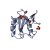
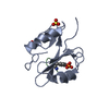
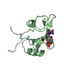
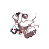
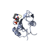
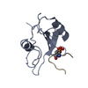
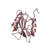
| Title | Structural Basis for Differential Recognition of Tyrosine Phosphorylated Sites in the Linker for Activation of T cells (LAT) by the Adaptor Protein Gads | ||||||
 Components Components |
| ||||||
 Keywords Keywords | PEPTIDE BINDING PROTEIN / SH2 / Gads / LAT / phosphopeptide | ||||||
| Function / homology |  Function and homology information Function and homology informationFLT3 Signaling / Co-stimulation by CD28 / FCERI mediated MAPK activation / Signaling by SCF-KIT / Generation of second messenger molecules / FCERI mediated Ca+2 mobilization / DAP12 signaling / regulation of MAPK cascade / phosphotyrosine residue binding / endosome ...FLT3 Signaling / Co-stimulation by CD28 / FCERI mediated MAPK activation / Signaling by SCF-KIT / Generation of second messenger molecules / FCERI mediated Ca+2 mobilization / DAP12 signaling / regulation of MAPK cascade / phosphotyrosine residue binding / endosome / signal transduction / nucleoplasm / nucleus / plasma membrane / cytoplasm / cytosol Similarity search - Function | ||||||
| Biological species |  | ||||||
| Method |  X-RAY DIFFRACTION / MOLECULAR REPLACEMENT / Resolution: 1.8 Å X-RAY DIFFRACTION / MOLECULAR REPLACEMENT / Resolution: 1.8 Å | ||||||
 Authors Authors | Cho, S. / Mariuzza, R.A. | ||||||
 Citation Citation | Journal: Embo J. / Year: 2004 Title: Structural basis for differential recognition of tyrosine-phosphorylated sites in the linker for activation of T cells (LAT) by the adaptor Gads. Authors: Cho, S. / Velikovsky, C.A. / Swaminathan, C.P. / Houtman, J.C. / Samelson, L.E. / Mariuzza, R.A. | ||||||
| History |
|
- Structure visualization
| Structure viewer | Molecule: MolmilJmol/JSmol |
|---|
- Downloads & links
-Download
| PDBx/mmCIF format | 1r1q.cif.gz | 61.3 KB | Display | PDBx/mmCIF format |
|---|---|---|---|---|
| PDB format | pdb1r1q.ent.gz | 44.8 KB | Display | PDB format |
| PDBx/mmJSON format | 1r1q.json.gz | Tree view | PDBx/mmJSON format | |
| Others |  Other downloads Other downloads |
-Validation report
| Arichive directory | https://data.pdbj.org/pub/pdb/validation_reports/r1/1r1qftp://data.pdbj.org/pub/pdb/validation_reports/r1/1r1q | HTTPS FTP |
|---|
-Related structure data


-Links
 PDBj
PDBj


- Assembly
| Deposited unit | 
| ||||||||
|---|---|---|---|---|---|---|---|---|---|
| 1 | 
| ||||||||
| 2 | 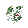
| ||||||||
| 3 | 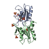
| ||||||||
| Unit cell |
|
-Components
| #1: Protein | Mass: 11823.300 Da / Num. of mol.: 2 / Fragment: Gads-SH2 domain Source method: isolated from a genetically manipulated source Source: (gene. exp.)  #2: Protein/peptide | Mass: 885.878 Da / Num. of mol.: 2 / Source method: obtained synthetically / Details: THE PEPTIDE WAS CHEMICALLY SYNTHESIZED #3: Chemical | ChemComp-SO4 /   Mass: 96.063 Da / Num. of mol.: 4 / Source method: obtained synthetically / Formula: SO4 Mass: 96.063 Da / Num. of mol.: 4 / Source method: obtained synthetically / Formula: SO4#4: Water | ChemComp-HOH / |  Mass: 18.015 Da / Num. of mol.: 142 / Source method: isolated from a natural source / Formula: H2O Mass: 18.015 Da / Num. of mol.: 142 / Source method: isolated from a natural source / Formula: H2OHas protein modification | Y | |
|---|
-Experimental details
-Experiment
| Experiment | Method: X-RAY DIFFRACTION / Number of used crystals: 1 |
|---|
- Sample preparation
| Crystal | Density Matthews: 1.88 Å3/Da / Density % sol: 34.43 % |
|---|---|
| Crystal grow | Temperature: 298 K / Method: vapor diffusion / pH: 8.7 Details: 0.1mM Tris-HCl, 2.5M ammonium sulfate, pH 8.7, VAPOR DIFFUSION, temperature 298K |
-Data collection
| Diffraction | Mean temperature: 100 K |
|---|---|
| Diffraction source | Source: ROTATING ANODE / Type: RIGAKU RU200 / Wavelength: 1.5418 Å |
| Detector | Type: RIGAKU RAXIS IV / Detector: IMAGE PLATE / Date: Apr 23, 2003 / Details: mirrors |
| Radiation | Monochromator: osmic mirror / Protocol: SINGLE WAVELENGTH / Monochromatic (M) / Laue (L): M / Scattering type: x-ray |
| Radiation wavelength | Wavelength: 1.5418 Å / Relative weight: 1 |
| Reflection | Resolution: 1.62→43.1 Å / Num. all: 117803 / Num. obs: 26632 / % possible obs: 96.6 % / Observed criterion σ(F): 2 / Observed criterion σ(I): 2 |
- Processing
| Software |
| ||||||||||||||||||||||||||||||||||||||||||||||||||||||||||||||||||||||||||||||||||||||||||||||||||||||||||||||||||||||||||||||||||
|---|---|---|---|---|---|---|---|---|---|---|---|---|---|---|---|---|---|---|---|---|---|---|---|---|---|---|---|---|---|---|---|---|---|---|---|---|---|---|---|---|---|---|---|---|---|---|---|---|---|---|---|---|---|---|---|---|---|---|---|---|---|---|---|---|---|---|---|---|---|---|---|---|---|---|---|---|---|---|---|---|---|---|---|---|---|---|---|---|---|---|---|---|---|---|---|---|---|---|---|---|---|---|---|---|---|---|---|---|---|---|---|---|---|---|---|---|---|---|---|---|---|---|---|---|---|---|---|---|---|---|---|
| Refinement | Method to determine structure: MOLECULAR REPLACEMENT / Resolution: 1.8→43.03 Å / Cor.coef. Fo:Fc: 0.954 / Cor.coef. Fo:Fc free: 0.939 / SU B: 3.007 / SU ML: 0.093 / Cross valid method: THROUGHOUT / σ(F): 2 / ESU R: 0.155 / ESU R Free: 0.143 / Stereochemistry target values: MAXIMUM LIKELIHOOD
| ||||||||||||||||||||||||||||||||||||||||||||||||||||||||||||||||||||||||||||||||||||||||||||||||||||||||||||||||||||||||||||||||||
| Solvent computation | Ion probe radii: 0.8 Å / Shrinkage radii: 0.8 Å / VDW probe radii: 1.4 Å / Solvent model: BABINET MODEL WITH MASK | ||||||||||||||||||||||||||||||||||||||||||||||||||||||||||||||||||||||||||||||||||||||||||||||||||||||||||||||||||||||||||||||||||
| Displacement parameters | Biso mean: 21.308 Å2
| ||||||||||||||||||||||||||||||||||||||||||||||||||||||||||||||||||||||||||||||||||||||||||||||||||||||||||||||||||||||||||||||||||
| Refinement step | Cycle: LAST / Resolution: 1.8→43.03 Å
| ||||||||||||||||||||||||||||||||||||||||||||||||||||||||||||||||||||||||||||||||||||||||||||||||||||||||||||||||||||||||||||||||||
| Refine LS restraints |
| ||||||||||||||||||||||||||||||||||||||||||||||||||||||||||||||||||||||||||||||||||||||||||||||||||||||||||||||||||||||||||||||||||
| LS refinement shell | Resolution: 1.8→1.847 Å / Total num. of bins used: 20 /
|
