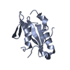
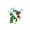
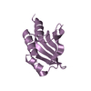
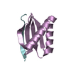
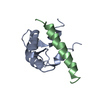
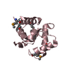
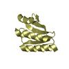
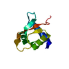
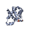
- PDB-3m03: Crystal structure of human Orc6 fragment -
+
Open data
ID or keywords:
Loading...
-
Basic information
Entry
Database: PDB / ID: 3m03
Title
Crystal structure of human Orc6 fragment
Components
Origin recognition complex subunit 6
Keywords
DNA BINDING PROTEIN / helix turn helix / Origin recognition complex / DNA replication
Function / homology
Function and homology information
CDC6 association with the ORC:origin complex / origin recognition complex / E2F-enabled inhibition of pre-replication complex formation / nuclear origin of replication recognition complex / Activation of the pre-replicative complex / DNA replication initiation / Activation of ATR in response to replication stress / Assembly of the ORC complex at the origin of replication / Assembly of the pre-replicative complex / fibrillar center ...CDC6 association with the ORC:origin complex / origin recognition complex / E2F-enabled inhibition of pre-replication complex formation / nuclear origin of replication recognition complex / Activation of the pre-replicative complex / DNA replication initiation / Activation of ATR in response to replication stress / Assembly of the ORC complex at the origin of replication / Assembly of the pre-replicative complex / fibrillar center / Orc1 removal from chromatin / DNA binding / nucleoplasm / membrane / nucleus / cytosol Similarity search - Function
Type: RIGAKU RAXIS IV / Detector: IMAGE PLATE / Date: Nov 1, 2009
Radiation
Protocol: SINGLE WAVELENGTH / Monochromatic (M) / Laue (L): M / Scattering type: x-ray
Radiation wavelength
Wavelength: 1.5418 Å / Relative weight: 1
Reflection
Resolution: 2.5→50 Å / Num. obs: 12005 / % possible obs: 99.9 % / Observed criterion σ(I): 2 / Redundancy: 7.1 % / Biso Wilson estimate: 57.8 Å2 / Rmerge(I) obs: 0.071 / Net I/σ(I): 38.9
Reflection shell
Resolution: 2.5→2.59 Å / Redundancy: 7.1 % / Rmerge(I) obs: 0.51 / Mean I/σ(I) obs: 4.9 / Num. unique all: 1226 / % possible all: 100
-
Processing
Software
Name
Version
Classification
CrystalClear
datacollection
SOLVE
phasing
REFMAC
5.5.0102
refinement
HKL-2000
datareduction
HKL-2000
datascaling
Refinement
Method to determine structure: SAD / Resolution: 2.5→50 Å / Cor.coef. Fo:Fc: 0.93 / Cor.coef. Fo:Fc free: 0.91 / SU B: 20.211 / SU ML: 0.21 / Cross valid method: THROUGHOUT / ESU R: 0.517 / ESU R Free: 0.296 / Stereochemistry target values: MAXIMUM LIKELIHOOD / Details: HYDROGENS HAVE BEEN ADDED IN THE RIDING POSITIONS
Rfactor
Num. reflection
% reflection
Selection details
Rfree
0.26618
572
4.8 %
RANDOM
Rwork
0.22599
-
-
-
obs
0.2279
11398
99.78 %
-
Solvent computation
Ion probe radii: 0.8 Å / Shrinkage radii: 0.8 Å / VDW probe radii: 1.4 Å / Solvent model: MASK
Displacement parameters
Biso mean: 58.183 Å2
Baniso -1
Baniso -2
Baniso -3
1-
-0.25 Å2
0 Å2
0 Å2
2-
-
-0.25 Å2
0 Å2
3-
-
-
0.5 Å2
Refinement step
Cycle: LAST / Resolution: 2.5→50 Å
Protein
Nucleic acid
Ligand
Solvent
Total
Num. atoms
2101
0
17
58
2176
Refine LS restraints
Refine-ID
Type
Dev ideal
Dev ideal target
Number
X-RAY DIFFRACTION
r_bond_refined_d
0.013
0.022
2130
X-RAY DIFFRACTION
r_angle_refined_deg
1.399
2.011
2849
X-RAY DIFFRACTION
r_dihedral_angle_1_deg
7.461
5
269
X-RAY DIFFRACTION
r_dihedral_angle_2_deg
37.226
25.89
73
X-RAY DIFFRACTION
r_dihedral_angle_3_deg
21.082
15
450
X-RAY DIFFRACTION
r_dihedral_angle_4_deg
14.268
15
9
X-RAY DIFFRACTION
r_chiral_restr
0.106
0.2
350
X-RAY DIFFRACTION
r_gen_planes_refined
0.01
0.02
1444
X-RAY DIFFRACTION
r_mcbond_it
1.107
1.5
1362
X-RAY DIFFRACTION
r_mcangle_it
1.968
2
2192
X-RAY DIFFRACTION
r_scbond_it
3.345
3
768
X-RAY DIFFRACTION
r_scangle_it
5.29
4.5
657
LS refinement shell
Resolution: 2.503→2.568 Å / Total num. of bins used: 20
Rfactor
Num. reflection
% reflection
Rfree
0.404
37
-
Rwork
0.313
854
-
obs
-
1226
100 %
Refinement TLS params.
Method: refined / Refine-ID: X-RAY DIFFRACTION
ID
L11 (°2)
L12 (°2)
L13 (°2)
L22 (°2)
L23 (°2)
L33 (°2)
S11 (Å °)
S12 (Å °)
S13 (Å °)
S21 (Å °)
S22 (Å °)
S23 (Å °)
S31 (Å °)
S32 (Å °)
S33 (Å °)
T11 (Å2)
T12 (Å2)
T13 (Å2)
T22 (Å2)
T23 (Å2)
T33 (Å2)
Origin x (Å)
Origin y (Å)
Origin z (Å)
1
4.5544
0.3267
0.9433
2.8044
-0.8621
4.8695
-0.138
0.1089
0.374
0.0916
-0.0657
-0.2669
-0.5397
0.7829
0.2037
0.1252
-0.1111
-0.0282
0.2716
0.0321
0.0853
32.0746
21.4964
28.1548
2
2.0816
0.382
0.1885
5.8403
0.2478
1.8924
-0.018
-0.016
-0.329
-0.0121
0.0186
0.0762
0.0472
0.2989
-0.0006
0.0154
0.0367
0.0221
0.1376
0.0436
0.1854
20.0729
0.2213
30.735
3
18.4373
-3.7986
2.6859
5.2872
0.2719
2.183
-0.2133
1.0534
-0.1351
0.2776
0.2661
0.0302
-0.2157
-0.1461
-0.0528
0.3118
0.0025
-0.0062
0.8047
0.0943
0.4312
58.8419
10.4321
33.4452
Refinement TLS group
ID
Refine-ID
Refine TLS-ID
Auth asym-ID
Auth seq-ID
1
X-RAY DIFFRACTION
1
A
94 - 186
2
X-RAY DIFFRACTION
2
B
93 - 187
3
X-RAY DIFFRACTION
3
C
97 - 185
+
About Yorodumi
-
News
-
Feb 9, 2022. New format data for meta-information of EMDB entries
New format data for meta-information of EMDB entries
Version 3 of the EMDB header file is now the official format.
The previous official version 1.9 will be removed from the archive.
In the structure databanks used in Yorodumi, some data are registered as the other names, "COVID-19 virus" and "2019-nCoV". Here are the details of the virus and the list of structure data.
Jan 31, 2019. EMDB accession codes are about to change! (news from PDBe EMDB page)
EMDB accession codes are about to change! (news from PDBe EMDB page)
The allocation of 4 digits for EMDB accession codes will soon come to an end. Whilst these codes will remain in use, new EMDB accession codes will include an additional digit and will expand incrementally as the available range of codes is exhausted. The current 4-digit format prefixed with “EMD-” (i.e. EMD-XXXX) will advance to a 5-digit format (i.e. EMD-XXXXX), and so on. It is currently estimated that the 4-digit codes will be depleted around Spring 2019, at which point the 5-digit format will come into force.
The EM Navigator/Yorodumi systems omit the EMD- prefix.
Related info.:Q: What is EMD? / ID/Accession-code notation in Yorodumi/EM Navigator
Yorodumi is a browser for structure data from EMDB, PDB, SASBDB, etc.
This page is also the successor to EM Navigator detail page, and also detail information page/front-end page for Omokage search.
The word "yorodu" (or yorozu) is an old Japanese word meaning "ten thousand". "mi" (miru) is to see.
Related info.:EMDB / PDB / SASBDB / Comparison of 3 databanks / Yorodumi Search / Aug 31, 2016. New EM Navigator & Yorodumi / Yorodumi Papers / Jmol/JSmol / Function and homology information / Changes in new EM Navigator and Yorodumi
 Movie
Movie Controller


 Open data
 Basic information
 Components
 Keywords
 Function and homology information
 Homo sapiens (human)
 X-RAY DIFFRACTION /
  Authors
 Citation
 Structure visualization
 Downloads & links
 Other downloads

 PDBj
PDBj


 Assembly


 Mass: 195.237 Da / Num. of mol.: 1 / Source method: obtained synthetically / Formula: C6H13NO4S / Comment: pH buffer*YM
 Mass: 96.063 Da / Num. of mol.: 1 / Source method: obtained synthetically / Formula: SO4
 Mass: 18.015 Da / Num. of mol.: 58 / Source method: isolated from a natural source / Formula: H2O
 Sample preparation
 Processing