 Movie
Movie Controller


+ Open data

- Basic information
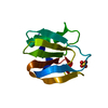
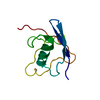
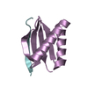
| Entry | Database: PDB / ID: 1hxr | ||||||
|---|---|---|---|---|---|---|---|
| Title | CRYSTAL STRUCTURE OF MSS4 AT 1.65 ANGSTROMS | ||||||
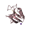
 Components Components | GUANINE NUCLEOTIDE EXCHANGE FACTOR MSS4 | ||||||
 Keywords Keywords | METAL BINDING PROTEIN / nucleotide exchange factor / Rab GTPase / membrane trafficking / Zn binding site | ||||||
| Function / homology |  Function and homology information Function and homology informationpost-Golgi vesicle-mediated transport / small GTPase-mediated signal transduction / guanyl-nucleotide exchange factor activity / protein transport / zinc ion binding / membrane / cytosol Similarity search - Function | ||||||
| Biological species |  | ||||||
| Method |  X-RAY DIFFRACTION / SIR/Molecular Replacement / Resolution: 1.65 Å X-RAY DIFFRACTION / SIR/Molecular Replacement / Resolution: 1.65 Å | ||||||
 Authors Authors | Zhu, Z. / Dumas, J.J. / Lietzke, S.E. / Lambright, D.G. | ||||||
 Citation Citation | Journal: Biochemistry / Year: 2001 Title: A helical turn motif in Mss4 is a critical determinant of Rab binding and nucleotide release. Authors: Zhu, Z. / Dumas, J.J. / Lietzke, S.E. / Lambright, D.G. | ||||||
| History |
|
- Structure visualization
| Structure viewer | Molecule: MolmilJmol/JSmol |
|---|
- Downloads & links
-Download
| PDBx/mmCIF format | 1hxr.cif.gz | 59.6 KB | Display | PDBx/mmCIF format |
|---|---|---|---|---|
| PDB format | pdb1hxr.ent.gz | 41.6 KB | Display | PDB format |
| PDBx/mmJSON format | 1hxr.json.gz | Tree view | PDBx/mmJSON format | |
| Others |  Other downloads Other downloads |
-Validation report
| Arichive directory | https://data.pdbj.org/pub/pdb/validation_reports/hx/1hxrftp://data.pdbj.org/pub/pdb/validation_reports/hx/1hxr | HTTPS FTP |
|---|
-Related structure data
| Related structure data | 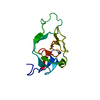 1fwqS S: Starting model for refinement |
|---|---|
| Similar structure data |


-Links
 PDBj
PDBj- Assembly
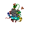
| Deposited unit | 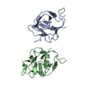
| ||||||||
|---|---|---|---|---|---|---|---|---|---|
| 1 | 
| ||||||||
| 2 | 
| ||||||||
| Unit cell |
|
-Components
| #1: Protein | Mass: 12999.825 Da / Num. of mol.: 2 Source method: isolated from a genetically manipulated source Source: (gene. exp.)  #2: Chemical |   Mass: 65.409 Da / Num. of mol.: 2 / Source method: obtained synthetically / Formula: Zn Mass: 65.409 Da / Num. of mol.: 2 / Source method: obtained synthetically / Formula: Zn#3: Water | ChemComp-HOH / |  Mass: 18.015 Da / Num. of mol.: 187 / Source method: isolated from a natural source / Formula: H2O Mass: 18.015 Da / Num. of mol.: 187 / Source method: isolated from a natural source / Formula: H2O |
|---|
-Experimental details
-Experiment
| Experiment | Method: X-RAY DIFFRACTION / Number of used crystals: 2 |
|---|
- Sample preparation
| Crystal | Density Matthews: 1.98 Å3/Da / Density % sol: 37.94 % | ||||||||||||||||||||||||||||||
|---|---|---|---|---|---|---|---|---|---|---|---|---|---|---|---|---|---|---|---|---|---|---|---|---|---|---|---|---|---|---|---|
| Crystal grow | Temperature: 277 K / Method: vapor diffusion, hanging drop / pH: 8 Details: 14% PEG 6000, 50 mM Tris, 50 mM Mg(OAc)2, 10% glycerol, pH 8.0, VAPOR DIFFUSION, HANGING DROP, temperature 277K | ||||||||||||||||||||||||||||||
| Crystal grow | *PLUS Temperature: 4 ℃ | ||||||||||||||||||||||||||||||
| Components of the solutions | *PLUS
|
-Data collection
| Diffraction | Mean temperature: 100 K |
|---|---|
| Diffraction source | Source: ROTATING ANODE / Type: RIGAKU RU200 / Wavelength: 1.5418 Å |
| Detector | Type: MARRESEARCH / Detector: IMAGE PLATE / Date: Jan 8, 1999 / Details: mirrors |
| Radiation | Protocol: SINGLE WAVELENGTH / Monochromatic (M) / Laue (L): M / Scattering type: x-ray |
| Radiation wavelength | Wavelength: 1.5418 Å / Relative weight: 1 |
| Reflection | Resolution: 1.65→20 Å / Num. all: 24609 / Num. obs: 24609 / % possible obs: 99.6 % / Observed criterion σ(I): -3 / Redundancy: 13 % / Biso Wilson estimate: 16.4 Å2 / Rsym value: 0.065 / Net I/σ(I): 17.1 |
| Reflection shell | Resolution: 1.65→7 Å / Redundancy: 3 % / Mean I/σ(I) obs: 3.7 / Num. unique all: 1993 / Rsym value: 0.282 / % possible all: 97.2 |
| Reflection | *PLUS Rmerge(I) obs: 0.065 |
| Reflection shell | *PLUS % possible obs: 97.2 % / Rmerge(I) obs: 0.282 |
- Processing
| Software |
| ||||||||||||||||||||||||||||||
|---|---|---|---|---|---|---|---|---|---|---|---|---|---|---|---|---|---|---|---|---|---|---|---|---|---|---|---|---|---|---|---|
| Refinement | Method to determine structure: SIR/Molecular Replacement Starting model: PDB entry 1FWQ Resolution: 1.65→7 Å / σ(F): 2 / Stereochemistry target values: Engh & Huber
| ||||||||||||||||||||||||||||||
| Refinement step | Cycle: LAST / Resolution: 1.65→7 Å
| ||||||||||||||||||||||||||||||
| Software | *PLUS Name: X-PLOR / Classification: refinement | ||||||||||||||||||||||||||||||
| Refine LS restraints | *PLUS
|
