 Movie
Movie Controller


+ Open data

- Basic information
| Entry | Database: PDB / ID: 2vpj | ||||||
|---|---|---|---|---|---|---|---|
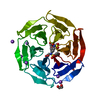
| Title | Crystal structure of the Kelch domain of human KLHL12 | ||||||
 Components Components | KELCH-LIKE PROTEIN 12 | ||||||
 Keywords Keywords | PROTEIN BINDING / ADAPTOR PROTEIN / WNT SIGNALING PATHWAY / PROTEIN-BINDING / UBIQUITIN DEGRADATION / UBL CONJUGATION PATHWAY / CUL3 / KELCH REPEAT / PHOSPHOPROTEIN / WNT SIGNALLING | ||||||
| Function / homology |  Function and homology information Function and homology informationneural crest formation / neural crest cell development / COPII vesicle coat assembly / COPII vesicle coat / COPII-coated ER to Golgi transport vesicle / Cul3-RING ubiquitin ligase complex / endoplasmic reticulum to Golgi vesicle-mediated transport / protein monoubiquitination / ubiquitin-like ligase-substrate adaptor activity / Degradation of DVL ...neural crest formation / neural crest cell development / COPII vesicle coat assembly / COPII vesicle coat / COPII-coated ER to Golgi transport vesicle / Cul3-RING ubiquitin ligase complex / endoplasmic reticulum to Golgi vesicle-mediated transport / protein monoubiquitination / ubiquitin-like ligase-substrate adaptor activity / Degradation of DVL / Wnt signaling pathway / centriolar satellite / proteasome-mediated ubiquitin-dependent protein catabolic process / identical protein binding / cytoplasm / cytosol Similarity search - Function | ||||||
| Biological species |  HOMO SAPIENS (human) HOMO SAPIENS (human) | ||||||
| Method |  X-RAY DIFFRACTION / SYNCHROTRON / MOLECULAR REPLACEMENT / Resolution: 1.85 Å X-RAY DIFFRACTION / SYNCHROTRON / MOLECULAR REPLACEMENT / Resolution: 1.85 Å | ||||||
 Authors Authors | Keates, T. / Pike, A.C.W. / Bullock, A.N. / Salah, E. / Filippakopoulos, P. / Roos, A.K. / von Delft, F. / Savitsky, P. / Weigelt, J. / Edwards, A. ...Keates, T. / Pike, A.C.W. / Bullock, A.N. / Salah, E. / Filippakopoulos, P. / Roos, A.K. / von Delft, F. / Savitsky, P. / Weigelt, J. / Edwards, A. / Arrowsmith, C.H. / Bountra, C. / Knapp, S. | ||||||
 Citation Citation | Journal: J.Biol.Chem. / Year: 2013 Title: Structural Basis for Cul3 Assembly with the Btb-Kelch Family of E3 Ubiquitin Ligases. Authors: Canning, P. / Cooper, C.D.O. / Krojer, T. / Murray, J.W. / Pike, A.C.W. / Chaikuad, A. / Keates, T. / Thangaratnarajah, C. / Hojzan, V. / Marsden, B.D. / Gileadi, O. / Knapp, S. / von Delft, F. / Bullock, A.N. | ||||||
| History |
|
- Structure visualization
| Structure viewer | Molecule: MolmilJmol/JSmol |
|---|
- Downloads & links
-Download
| PDBx/mmCIF format | 2vpj.cif.gz | 71.5 KB | Display | PDBx/mmCIF format |
|---|---|---|---|---|
| PDB format | pdb2vpj.ent.gz | 51.8 KB | Display | PDB format |
| PDBx/mmJSON format | 2vpj.json.gz | Tree view | PDBx/mmJSON format | |
| Others |  Other downloads Other downloads |
-Validation report
| Arichive directory | https://data.pdbj.org/pub/pdb/validation_reports/vp/2vpjftp://data.pdbj.org/pub/pdb/validation_reports/vp/2vpj | HTTPS FTP |
|---|
-Related structure data
| Related structure data |  2xn4C  3ii7C  4ap2C  4apfC 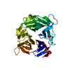 4ascC  2dyhS S: Starting model for refinement C: citing same article ( |
|---|---|
| Similar structure data |


-Links
 PDBj
PDBj


- Assembly
| Deposited unit | 
| ||||||||
|---|---|---|---|---|---|---|---|---|---|
| 1 |
| ||||||||
| Unit cell |
|
-Components
| #1: Protein | Mass: 32905.879 Da / Num. of mol.: 1 / Fragment: KELCH DOMAIN, RESIDUES 268-567 Source method: isolated from a genetically manipulated source Source: (gene. exp.) HOMO SAPIENS (human) / Plasmid: PNIC28-BSA4 / Production host:  | ||
|---|---|---|---|
| #2: Chemical |   Mass: 59.044 Da / Num. of mol.: 2 / Source method: obtained synthetically / Formula: C2H3O2 Mass: 59.044 Da / Num. of mol.: 2 / Source method: obtained synthetically / Formula: C2H3O2#3: Water | ChemComp-HOH / |  Mass: 18.015 Da / Num. of mol.: 132 / Source method: isolated from a natural source / Formula: H2O Mass: 18.015 Da / Num. of mol.: 132 / Source method: isolated from a natural source / Formula: H2O |
-Experimental details
-Experiment
| Experiment | Method: X-RAY DIFFRACTION / Number of used crystals: 1 |
|---|
- Sample preparation
| Crystal | Density Matthews: 1.77 Å3/Da / Density % sol: 30.42 % / Description: NONE |
|---|---|
| Crystal grow | pH: 4.6 Details: 0.2M AMMONIUM ACETATE, 0.1M SODIUM ACETATE PH4.6, 30% PEG4K |
-Data collection
| Diffraction | Mean temperature: 100 K |
|---|---|
| Diffraction source | Source: SYNCHROTRON / Site: SLS  / Beamline: X10SA / Wavelength: 0.98068 / Beamline: X10SA / Wavelength: 0.98068 |
| Detector | Type: MARRESEARCH / Detector: CCD / Date: Jan 26, 2008 |
| Radiation | Protocol: SINGLE WAVELENGTH / Monochromatic (M) / Laue (L): M / Scattering type: x-ray |
| Radiation wavelength | Wavelength: 0.98068 Å / Relative weight: 1 |
| Reflection | Resolution: 1.85→42.3 Å / Num. obs: 19527 / % possible obs: 99.4 % / Observed criterion σ(I): 0 / Redundancy: 4.3 % / Biso Wilson estimate: 23.09 Å2 / Rmerge(I) obs: 0.09 / Net I/σ(I): 10.6 |
| Reflection shell | Resolution: 1.85→1.95 Å / Redundancy: 3.4 % / Rmerge(I) obs: 0.63 / Mean I/σ(I) obs: 2 / % possible all: 96.1 |
- Processing
| Software |
| ||||||||||||||||||||||||||||||||||||||||||||||||||||||||||||||||||||||||||||||||||||||||||||||||||||||||||||||||||||||||||||||||||||||||||||||||||||||||||||||||||||||||||||||||||||||
|---|---|---|---|---|---|---|---|---|---|---|---|---|---|---|---|---|---|---|---|---|---|---|---|---|---|---|---|---|---|---|---|---|---|---|---|---|---|---|---|---|---|---|---|---|---|---|---|---|---|---|---|---|---|---|---|---|---|---|---|---|---|---|---|---|---|---|---|---|---|---|---|---|---|---|---|---|---|---|---|---|---|---|---|---|---|---|---|---|---|---|---|---|---|---|---|---|---|---|---|---|---|---|---|---|---|---|---|---|---|---|---|---|---|---|---|---|---|---|---|---|---|---|---|---|---|---|---|---|---|---|---|---|---|---|---|---|---|---|---|---|---|---|---|---|---|---|---|---|---|---|---|---|---|---|---|---|---|---|---|---|---|---|---|---|---|---|---|---|---|---|---|---|---|---|---|---|---|---|---|---|---|---|---|
| Refinement | Method to determine structure: MOLECULAR REPLACEMENT Starting model: PDB ENTRY 2DYH Resolution: 1.85→42.3 Å / Cor.coef. Fo:Fc: 0.972 / Cor.coef. Fo:Fc free: 0.947 / SU B: 8.481 / SU ML: 0.122 / TLS residual ADP flag: LIKELY RESIDUAL / Cross valid method: THROUGHOUT / ESU R: 0.153 / ESU R Free: 0.149 / Stereochemistry target values: MAXIMUM LIKELIHOOD / Details: HYDROGENS HAVE BEEN ADDED IN THE RIDING POSITIONS.
| ||||||||||||||||||||||||||||||||||||||||||||||||||||||||||||||||||||||||||||||||||||||||||||||||||||||||||||||||||||||||||||||||||||||||||||||||||||||||||||||||||||||||||||||||||||||
| Solvent computation | Ion probe radii: 0.8 Å / Shrinkage radii: 0.8 Å / VDW probe radii: 1.2 Å / Solvent model: MASK | ||||||||||||||||||||||||||||||||||||||||||||||||||||||||||||||||||||||||||||||||||||||||||||||||||||||||||||||||||||||||||||||||||||||||||||||||||||||||||||||||||||||||||||||||||||||
| Displacement parameters | Biso mean: 23.13 Å2
| ||||||||||||||||||||||||||||||||||||||||||||||||||||||||||||||||||||||||||||||||||||||||||||||||||||||||||||||||||||||||||||||||||||||||||||||||||||||||||||||||||||||||||||||||||||||
| Refinement step | Cycle: LAST / Resolution: 1.85→42.3 Å
| ||||||||||||||||||||||||||||||||||||||||||||||||||||||||||||||||||||||||||||||||||||||||||||||||||||||||||||||||||||||||||||||||||||||||||||||||||||||||||||||||||||||||||||||||||||||
| Refine LS restraints |
|