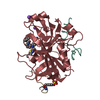
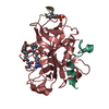
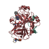
[English] 日本語
Yorodumi- PDB-2dyh: Crystal structure of the Keap1 protein in complexed with the N-te... -
+Open data
-Basic information
Entry | Database: PDB / ID: 2dyh | ||||||
---|---|---|---|---|---|---|---|
Title | Crystal structure of the Keap1 protein in complexed with the N-terminal region of the Nrf2 transcription factor | ||||||
Components |
| ||||||
Keywords | TRANSCRIPTION / bet-propeller / Kelch motif / Structural Genomics / NPPSFA / National Project on Protein Structural and Functional Analyses / RIKEN Structural Genomics/Proteomics Initiative / RSGI | ||||||
Function / homology | Function and homology information positive regulation of glutathione biosynthetic process / aflatoxin catabolic process / negative regulation of hematopoietic stem cell differentiation / Nuclear events mediated by NFE2L2 / GSK3B and BTRC:CUL1-mediated-degradation of NFE2L2 / negative regulation of cellular response to hypoxia / PERK-mediated unfolded protein response / cellular response to carbohydrate stimulus / negative regulation of vascular associated smooth muscle cell migration / regulation of removal of superoxide radicals ...positive regulation of glutathione biosynthetic process / aflatoxin catabolic process / negative regulation of hematopoietic stem cell differentiation / Nuclear events mediated by NFE2L2 / GSK3B and BTRC:CUL1-mediated-degradation of NFE2L2 / negative regulation of cellular response to hypoxia / PERK-mediated unfolded protein response / cellular response to carbohydrate stimulus / negative regulation of vascular associated smooth muscle cell migration / regulation of removal of superoxide radicals / KEAP1-NFE2L2 pathway / regulation of epidermal cell differentiation / Neddylation / cellular response to laminar fluid shear stress / Antigen processing: Ubiquitination & Proteasome degradation / Ub-specific processing proteases / cellular response to fluid shear stress / cellular response to angiotensin / negative regulation of response to oxidative stress / negative regulation of cardiac muscle cell apoptotic process / Cul3-RING ubiquitin ligase complex / proteasomal ubiquitin-independent protein catabolic process / regulation of innate immune response / regulation of embryonic development / transcription factor binding / ubiquitin-like ligase-substrate adaptor activity / centriolar satellite / positive regulation of blood vessel endothelial cell migration / positive regulation of blood coagulation / cellular response to interleukin-4 / cellular response to glucose starvation / negative regulation of endothelial cell apoptotic process / negative regulation of oxidative stress-induced intrinsic apoptotic signaling pathway / endoplasmic reticulum unfolded protein response / inclusion body / cellular response to copper ion / response to endoplasmic reticulum stress / : / molecular condensate scaffold activity / cell redox homeostasis / regulation of autophagy / transcription coregulator binding / response to ischemia / positive regulation of glucose import / actin filament / protein-DNA complex / adherens junction / positive regulation of neuron projection development / cellular response to hydrogen peroxide / RNA polymerase II transcription regulator complex / positive regulation of angiogenesis / positive regulation of reactive oxygen species metabolic process / disordered domain specific binding / cellular response to xenobiotic stimulus / cellular response to tumor necrosis factor / cellular response to oxidative stress / midbody / cellular response to hypoxia / ubiquitin-dependent protein catabolic process / DNA-binding transcription activator activity, RNA polymerase II-specific / proteasome-mediated ubiquitin-dependent protein catabolic process / in utero embryonic development / RNA polymerase II-specific DNA-binding transcription factor binding / transcription regulator complex / sequence-specific DNA binding / transcription cis-regulatory region binding / DNA-binding transcription factor activity, RNA polymerase II-specific / protein ubiquitination / inflammatory response / RNA polymerase II cis-regulatory region sequence-specific DNA binding / DNA-binding transcription factor activity / protein domain specific binding / negative regulation of gene expression / focal adhesion / centrosome / ubiquitin protein ligase binding / positive regulation of gene expression / regulation of DNA-templated transcription / chromatin / regulation of transcription by RNA polymerase II / positive regulation of DNA-templated transcription / Golgi apparatus / endoplasmic reticulum / positive regulation of transcription by RNA polymerase II / protein-containing complex / DNA binding / nucleoplasm / identical protein binding / nucleus / plasma membrane / cytoplasm / cytosol Similarity search - Function | ||||||
Biological species | Mus musculus (house mouse) | ||||||
Method | X-RAY DIFFRACTION / MOLECULAR REPLACEMENT / Resolution: 1.9 Å | ||||||
Authors | Padmanabhan, B. / Yokoyama, S. / RIKEN Structural Genomics/Proteomics Initiative (RSGI) | ||||||
Citation | Journal: Mol.Cell.Biol. / Year: 2007 Title: Different electrostatic potentials define ETGE and DLG motifs as hinge and latch in oxidative stress response Authors: Tong, K.I. / Padmanabhan, B. / Kobayashi, A. / Shang, C. / Hirotsu, Y. / Yokoyama, S. / Yamamoto, M. | ||||||
History |
|
-Structure visualization
Structure viewer | Molecule: MolmilJmol/JSmol |
---|
-Downloads & links
-Download
PDBx/mmCIF format | 2dyh.cif.gz | 82 KB | Display | PDBx/mmCIF format |
---|---|---|---|---|
PDB format | pdb2dyh.ent.gz | 59.8 KB | Display | PDB format |
PDBx/mmJSON format | 2dyh.json.gz | Tree view | PDBx/mmJSON format | |
Others | Other downloads |
-Validation report
Summary document | 2dyh_validation.pdf.gz | 384.9 KB | Display | wwPDB validaton report |
---|---|---|---|---|
Full document | 2dyh_full_validation.pdf.gz | 388.5 KB | Display | |
Data in XML | 2dyh_validation.xml.gz | 8.1 KB | Display | |
Data in CIF | 2dyh_validation.cif.gz | 13.6 KB | Display | |
Arichive directory | https://data.pdbj.org/pub/pdb/validation_reports/dy/2dyh ftp://data.pdbj.org/pub/pdb/validation_reports/dy/2dyh | HTTPS FTP |
-Related structure data
Related structure data | 1x2jS S: Starting model for refinement |
---|---|
Similar structure data | |
Other databases |
-Links
-Assembly
Deposited unit |
| ||||||||
---|---|---|---|---|---|---|---|---|---|
1 |
| ||||||||
Unit cell |
|
-Components
#1: Protein | Mass: 35006.277 Da / Num. of mol.: 1 / Fragment: Keap1-DC Source method: isolated from a genetically manipulated source Source: (gene. exp.) Mus musculus (house mouse) / Plasmid: pET15b / Production host: Escherichia coli (E. coli) / Strain (production host): BL21 CodonPlus (DE3)_RIL / References: UniProt: Q9Z2X8 | ||
---|---|---|---|
#2: Protein/peptide | Mass: 1801.053 Da / Num. of mol.: 1 / Source method: obtained synthetically / Details: Nrf2 peptide, synthetic peptide / References: UniProt: Q60795 | ||
#3: Chemical | ChemComp-SO4 / #4: Water | ChemComp-HOH / | |
-Experimental details
-Experiment
Experiment | Method: X-RAY DIFFRACTION / Number of used crystals: 1 |
---|
-Sample preparation
Crystal | Density Matthews: 2.34 Å3/Da / Density % sol: 47.45 % |
---|---|
Crystal grow | Temperature: 298 K / Method: vapor diffusion, sitting drop / pH: 5.6 Details: Ammonium sulfate, Lithium Sulfate, Sodium Citrate, pH 5.6, VAPOR DIFFUSION, SITTING DROP, temperature 298K |
-Data collection
Diffraction | Mean temperature: 103 K |
---|---|
Diffraction source | Source: ROTATING ANODE / Type: RIGAKU / Wavelength: 1.5418 Å |
Detector | Type: RIGAKU RAXIS IV++ / Detector: IMAGE PLATE / Date: Dec 16, 2005 / Details: mirrors |
Radiation | Protocol: SINGLE WAVELENGTH / Monochromatic (M) / Laue (L): M / Scattering type: x-ray |
Radiation wavelength | Wavelength: 1.5418 Å / Relative weight: 1 |
Reflection | Resolution: 1.9→50 Å / Num. all: 27043 / Num. obs: 26542 / % possible obs: 98.2 % / Observed criterion σ(I): -3 / Redundancy: 8.6 % / Rmerge(I) obs: 0.086 |
Reflection shell | Resolution: 1.9→1.97 Å / Redundancy: 3.6 % / Rmerge(I) obs: 0.398 / Num. unique all: 2359 / % possible all: 88 |
-Processing
Software |
| ||||||||||||||||||||||||||||||||||||||||||||||||||||||||||||||||||||||||||||||||||||||||||
---|---|---|---|---|---|---|---|---|---|---|---|---|---|---|---|---|---|---|---|---|---|---|---|---|---|---|---|---|---|---|---|---|---|---|---|---|---|---|---|---|---|---|---|---|---|---|---|---|---|---|---|---|---|---|---|---|---|---|---|---|---|---|---|---|---|---|---|---|---|---|---|---|---|---|---|---|---|---|---|---|---|---|---|---|---|---|---|---|---|---|---|
Refinement | Method to determine structure: MOLECULAR REPLACEMENT Starting model: PDB ENTRY 1X2J Resolution: 1.9→20 Å / Cor.coef. Fo:Fc: 0.965 / Cor.coef. Fo:Fc free: 0.956 / SU B: 3.172 / SU ML: 0.094 / Cross valid method: THROUGHOUT / σ(F): 0 / ESU R: 0.144 / ESU R Free: 0.134 / Stereochemistry target values: MAXIMUM LIKELIHOOD
| ||||||||||||||||||||||||||||||||||||||||||||||||||||||||||||||||||||||||||||||||||||||||||
Solvent computation | Ion probe radii: 0.8 Å / Shrinkage radii: 0.8 Å / VDW probe radii: 1.2 Å / Solvent model: BABINET MODEL WITH MASK | ||||||||||||||||||||||||||||||||||||||||||||||||||||||||||||||||||||||||||||||||||||||||||
Displacement parameters | Biso mean: 34.302 Å2
| ||||||||||||||||||||||||||||||||||||||||||||||||||||||||||||||||||||||||||||||||||||||||||
Refinement step | Cycle: LAST / Resolution: 1.9→20 Å
| ||||||||||||||||||||||||||||||||||||||||||||||||||||||||||||||||||||||||||||||||||||||||||
Refine LS restraints |
| ||||||||||||||||||||||||||||||||||||||||||||||||||||||||||||||||||||||||||||||||||||||||||
LS refinement shell | Resolution: 1.9→1.949 Å / Total num. of bins used: 20
|