 Movie
Movie Controller

[English] 日本語
 Yorodumi
- PDB-2qkb: Human RNase H catalytic domain mutant D210N in complex with 20-me... -
+ Open data

- Basic information
| Entry | Database: PDB / ID: 2qkb | ||||||
|---|---|---|---|---|---|---|---|
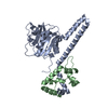
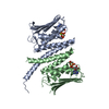
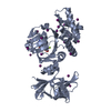
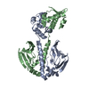
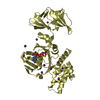
| Title | Human RNase H catalytic domain mutant D210N in complex with 20-mer RNA/DNA hybrid | ||||||
 Components Components |
| ||||||
 Keywords Keywords | HYDROLASE/DNA/RNA / RNase H / RNA/DNA hybrid / HYDROLASE-DNA-RNA COMPLEX | ||||||
| Function / homology |  Function and homology information Function and homology informationStrand-asynchronous mitochondrial DNA replication / DNA replication, removal of RNA primer / RNA catabolic process / ribonuclease H / RNA nuclease activity / RNA-DNA hybrid ribonuclease activity / nucleic acid binding / mitochondrial matrix / magnesium ion binding / mitochondrion / RNA binding Similarity search - Function | ||||||
| Biological species |  Homo sapiens (human) Homo sapiens (human) | ||||||
| Method |  X-RAY DIFFRACTION / SYNCHROTRON / MOLECULAR REPLACEMENT / Resolution: 2.4 Å X-RAY DIFFRACTION / SYNCHROTRON / MOLECULAR REPLACEMENT / Resolution: 2.4 Å | ||||||
 Authors Authors | Nowotny, M. / Gaidamakov, S.A. / Ghirlando, R. / Cerritelli, S.M. / Crouch, R.J. / Yang, W. | ||||||
 Citation Citation | Journal: Mol.Cell / Year: 2007 Title: Structure of Human RNase H1 Complexed with an RNA/DNA Hybrid: Insight into HIV Reverse Transcription Authors: Nowotny, M. / Gaidamakov, S.A. / Ghirlando, R. / Cerritelli, S.M. / Crouch, R.J. / Yang, W. | ||||||
| History |
|
- Structure visualization
| Structure viewer | Molecule: MolmilJmol/JSmol |
|---|
- Downloads & links
-Download
| PDBx/mmCIF format | 2qkb.cif.gz | 99.1 KB | Display | PDBx/mmCIF format |
|---|---|---|---|---|
| PDB format | pdb2qkb.ent.gz | 71.7 KB | Display | PDB format |
| PDBx/mmJSON format | 2qkb.json.gz | Tree view | PDBx/mmJSON format | |
| Others |  Other downloads Other downloads |
-Validation report
| Arichive directory | https://data.pdbj.org/pub/pdb/validation_reports/qk/2qkbftp://data.pdbj.org/pub/pdb/validation_reports/qk/2qkb | HTTPS FTP |
|---|
-Related structure data
| Related structure data | 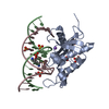 2qk9SC  2qkkC S: Starting model for refinement C: citing same article ( |
|---|---|
| Similar structure data |


-Links
 PDBj
PDBj


- Assembly
| Deposited unit | 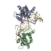
| ||||||||
|---|---|---|---|---|---|---|---|---|---|
| 1 |
| ||||||||
| Unit cell |
|
-Components
-RNA chain / DNA chain / Protein , 3 types, 4 molecules CDAB
| #1: RNA chain | Mass: 6382.854 Da / Num. of mol.: 1 / Source method: obtained synthetically |
|---|---|
| #2: DNA chain | Mass: 6149.978 Da / Num. of mol.: 1 / Source method: obtained synthetically |
| #3: Protein | Mass: 17291.861 Da / Num. of mol.: 2 / Fragment: C-terminal domain (residues 134-286) / Mutation: D210N Source method: isolated from a genetically manipulated source Source: (gene. exp.) Homo sapiens (human) / Gene: RNASEH1, RNH1 / Plasmid: pET15 / Species (production host): Escherichia coli / Production host:  |
-Non-polymers , 3 types, 62 molecules 


| #4: Chemical |  Mass: 96.063 Da / Num. of mol.: 2 / Source method: obtained synthetically / Formula: SO4 Mass: 96.063 Da / Num. of mol.: 2 / Source method: obtained synthetically / Formula: SO4#5: Chemical |  Mass: 62.068 Da / Num. of mol.: 2 / Source method: obtained synthetically / Formula: C2H6O2 Mass: 62.068 Da / Num. of mol.: 2 / Source method: obtained synthetically / Formula: C2H6O2#6: Water | ChemComp-HOH / | Mass: 18.015 Da / Num. of mol.: 58 / Source method: isolated from a natural source / Formula: H2O |
|---|
-Details
| Has protein modification | Y |
|---|
-Experimental details
-Experiment
| Experiment | Method: X-RAY DIFFRACTION / Number of used crystals: 1 |
|---|
- Sample preparation
| Crystal | Density Matthews: 2.49 Å3/Da / Density % sol: 50.63 % | ||||||||||||||||||||||||||||
|---|---|---|---|---|---|---|---|---|---|---|---|---|---|---|---|---|---|---|---|---|---|---|---|---|---|---|---|---|---|
| Crystal grow | Temperature: 294 K / Method: vapor diffusion, hanging drop / pH: 8.5 Details: 15% PEG 3350, 0.1 M LiSO4, 0.1 M Tris, pH 8.5, VAPOR DIFFUSION, HANGING DROP, temperature 294K | ||||||||||||||||||||||||||||
| Components of the solutions |
|
-Data collection
| Diffraction | Mean temperature: 100 K |
|---|---|
| Diffraction source | Source: SYNCHROTRON / Site: APS  / Beamline: 22-ID / Wavelength: 1 Å / Beamline: 22-ID / Wavelength: 1 Å |
| Radiation | Protocol: SINGLE WAVELENGTH / Monochromatic (M) / Laue (L): M / Scattering type: x-ray |
| Radiation wavelength | Wavelength: 1 Å / Relative weight: 1 |
| Reflection | Resolution: 2.4→40 Å / Num. all: 18670 / Num. obs: 16949 / % possible obs: 90.8 % / Rmerge(I) obs: 0.06 / Net I/σ(I): 35.8 |
| Reflection shell | Resolution: 2.4→2.49 Å / Rmerge(I) obs: 0.452 / Mean I/σ(I) obs: 2.7 |
- Processing
| Software |
| ||||||||||||||||||||||||||||||||||||
|---|---|---|---|---|---|---|---|---|---|---|---|---|---|---|---|---|---|---|---|---|---|---|---|---|---|---|---|---|---|---|---|---|---|---|---|---|---|
| Refinement | Method to determine structure: MOLECULAR REPLACEMENT Starting model: 2QK9 Resolution: 2.4→40 Å / Cross valid method: FREE R-VALUE / Stereochemistry target values: Engh & Huber Details: The last round of refinement in CNS was carried out with the pucker restraints for the DNA strand removed
| ||||||||||||||||||||||||||||||||||||
| Displacement parameters | Biso mean: 59.3 Å2 | ||||||||||||||||||||||||||||||||||||
| Refinement step | Cycle: LAST / Resolution: 2.4→40 Å
| ||||||||||||||||||||||||||||||||||||
| Refine LS restraints |
|
