Mass: 18.015 Da / Num. of mol.: 109 / Source method: isolated from a natural source / Formula: H2O
-
Experimental details
-
Experiment
Experiment
Method: X-RAY DIFFRACTION / Number of used crystals: 1
-
Sample preparation
Crystal grow
Temperature: 294 K / Method: vapor diffusion, hanging drop / pH: 5.6 Details: 1.5 M ammonium sulfate, 0.1 M Na citrate (pH 5.6) 20mM ATP or 3% 1,6-diaminohexane, VAPOR DIFFUSION, HANGING DROP, temperature 294K
Components of the solutions
ID
Name
Crystal-ID
Sol-ID
1
(NH4)2SO4
1
1
2
sodiumcitrate
1
1
3
1,6-diaminohexane
1
1
4
(NH4)2SO4
1
2
5
sodiumcitrate
1
2
6
1,6-diaminohexane
1
2
-
Data collection
Diffraction
Mean temperature: 100 K
Diffraction source
Source: SYNCHROTRON / Site: APS / Beamline: 22-ID
Detector
Type: MARRESEARCH / Detector: CCD
Radiation
Protocol: SINGLE WAVELENGTH / Monochromatic (M) / Laue (L): M / Scattering type: x-ray
Radiation wavelength
Relative weight: 1
Reflection
Resolution: 2.55→30 Å / Num. all: 22505 / Num. obs: 21164 / % possible obs: 94 % / Rmerge(I) obs: 0.074 / Net I/σ(I): 38.7
Reflection shell
Resolution: 2.55→2.64 Å / Rmerge(I) obs: 0.456 / Mean I/σ(I) obs: 4.1
-
Processing
Software
Name
Version
Classification
CNS
1.1
refinement
HKL-2000
datareduction
HKL-2000
datascaling
Refinement
Method to determine structure: MOLECULAR REPLACEMENT Starting model: Original low resolution strcture solved with the same crystal and SeMet SIRAS Resolution: 2.55→30 Å / Stereochemistry target values: Engh & Huber Details: The last round of refinement in CNS was carried out with the pucker restraints for the DNA strand removed
Rfactor
Num. reflection
% reflection
Selection details
Rfree
0.216
2085
-
Random
Rwork
0.19
-
-
-
all
-
22505
-
-
obs
-
21164
94 %
-
Displacement parameters
Biso mean: 43.3 Å2
Refinement step
Cycle: LAST / Resolution: 2.55→30 Å
Protein
Nucleic acid
Ligand
Solvent
Total
Num. atoms
1188
746
69
109
2112
Refine LS restraints
Refine-ID
Type
Dev ideal
Dev ideal target
X-RAY DIFFRACTION
c_bond_d
0.013483
X-RAY DIFFRACTION
c_angle_deg
1.86942
X-RAY DIFFRACTION
c_dihedral_angle_d
X-RAY DIFFRACTION
c_improper_angle_d
X-RAY DIFFRACTION
c_mcbond_it
1.418
1.5
X-RAY DIFFRACTION
c_mcangle_it
2.437
2
X-RAY DIFFRACTION
c_scbond_it
1.904
2
X-RAY DIFFRACTION
c_scangle_it
2.908
2.5
+
About Yorodumi
-
News
-
Feb 9, 2022. New format data for meta-information of EMDB entries
New format data for meta-information of EMDB entries
Version 3 of the EMDB header file is now the official format.
The previous official version 1.9 will be removed from the archive.
In the structure databanks used in Yorodumi, some data are registered as the other names, "COVID-19 virus" and "2019-nCoV". Here are the details of the virus and the list of structure data.
Jan 31, 2019. EMDB accession codes are about to change! (news from PDBe EMDB page)
EMDB accession codes are about to change! (news from PDBe EMDB page)
The allocation of 4 digits for EMDB accession codes will soon come to an end. Whilst these codes will remain in use, new EMDB accession codes will include an additional digit and will expand incrementally as the available range of codes is exhausted. The current 4-digit format prefixed with “EMD-” (i.e. EMD-XXXX) will advance to a 5-digit format (i.e. EMD-XXXXX), and so on. It is currently estimated that the 4-digit codes will be depleted around Spring 2019, at which point the 5-digit format will come into force.
The EM Navigator/Yorodumi systems omit the EMD- prefix.
Related info.:Q: What is EMD? / ID/Accession-code notation in Yorodumi/EM Navigator
Yorodumi is a browser for structure data from EMDB, PDB, SASBDB, etc.
This page is also the successor to EM Navigator detail page, and also detail information page/front-end page for Omokage search.
The word "yorodu" (or yorozu) is an old Japanese word meaning "ten thousand". "mi" (miru) is to see.
Related info.:EMDB / PDB / SASBDB / Comparison of 3 databanks / Yorodumi Search / Aug 31, 2016. New EM Navigator & Yorodumi / Yorodumi Papers / Jmol/JSmol / Function and homology information / Changes in new EM Navigator and Yorodumi
 Movie
Movie Controller

 Yorodumi
 Open data
 Basic information
 Components
 Keywords
 Function and homology information
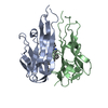
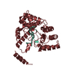
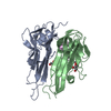
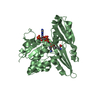
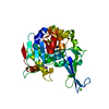
 Homo sapiens (human)
 X-RAY DIFFRACTION /
  Authors
 Citation
 Structure visualization
 Downloads & links
 Other downloads


 PDBj
PDBj


 Assembly
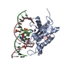


 Mass: 96.063 Da / Num. of mol.: 7 / Source method: obtained synthetically / Formula: SO4
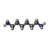
 Mass: 116.205 Da / Num. of mol.: 1 / Source method: obtained synthetically / Formula: C6H16N2
 Mass: 22.990 Da / Num. of mol.: 1 / Source method: obtained synthetically / Formula: Na
 Mass: 189.100 Da / Num. of mol.: 1 / Source method: obtained synthetically / Formula: C6H5O7
 Mass: 92.094 Da / Num. of mol.: 2 / Source method: obtained synthetically / Formula: C3H8O3
 Sample preparation
 / Beamline: 22-ID
 Processing