- PDB-5epv: Histidine kinase domain from the LOV-HK blue-light receptor from ... -
+
Open data
ID or keywords:
Loading...
-
Basic information
Entry
Database: PDB / ID: 5epv
Title
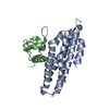
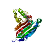
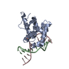
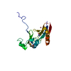
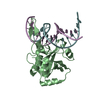
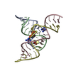
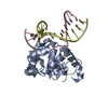
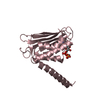
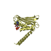
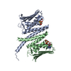
Histidine kinase domain from the LOV-HK blue-light receptor from Brucella abortus
Components
Blue-light-activated histidine kinase
Keywords
TRANSFERASE / Histidine kinase / HWE family / S-SAD phasing / multi-crystal data collection
Function / homology
Function and homology information
protein histidine kinase activity / histidine kinase / photoreceptor activity / ATP binding Similarity search - Function
Signal transduction histidine kinase, HWE region / HWE histidine kinase / HWE histidine kinase / PAS fold-3 / PAS fold / PAS domain / PAS-associated, C-terminal / PAC domain profile. / PAC motif / Motif C-terminal to PAS motifs (likely to contribute to PAS structural domain) ...Signal transduction histidine kinase, HWE region / HWE histidine kinase / HWE histidine kinase / PAS fold-3 / PAS fold / PAS domain / PAS-associated, C-terminal / PAC domain profile. / PAC motif / Motif C-terminal to PAS motifs (likely to contribute to PAS structural domain) / PAS domain / PAS repeat profile. / PAS domain / PAS domain superfamily Similarity search - Domain/homology
#1: Journal: Acta Crystallogr. D Biol. Crystallogr. / Year: 2015 Title: S-SAD phasing of monoclinic histidine kinase from Brucella abortus combining data from multiple crystals and orientations: an example of data-collection strategy and a posteriori analysis of ...Title: S-SAD phasing of monoclinic histidine kinase from Brucella abortus combining data from multiple crystals and orientations: an example of data-collection strategy and a posteriori analysis of different data combinations. Authors: Klinke, S. / Foos, N. / Rinaldi, J.J. / Paris, G. / Goldbaum, F.A. / Legrand, P. / Guimaraes, B.G. / Thompson, A.
Resolution: 2.51→50 Å / Num. all: 32760 / Num. obs: 32760 / % possible obs: 97.7 % / Redundancy: 3.8 % / Biso Wilson estimate: 72 Å2 / Rmerge(I) obs: 0.06 / Rsym value: 0.06 / Net I/σ(I): 14.3
Reflection shell
Resolution: 2.51→2.66 Å / Redundancy: 3.8 % / Rmerge(I) obs: 0.591 / Mean I/σ(I) obs: 2 / % possible all: 95.1
-
Processing
Software
Name
Version
Classification
MxCuBE
datacollection
XDS
datascaling
PHASER
phasing
SHELX
phasing
MOLREP
phasing
BUSTER
2.10.0
refinement
Coot
modelbuilding
XDS
datareduction
Refinement
Method to determine structure: SAD / Resolution: 2.51→48.55 Å / Cor.coef. Fo:Fc: 0.9433 / Cor.coef. Fo:Fc free: 0.9189 / SU R Cruickshank DPI: 0.39 / Cross valid method: THROUGHOUT / σ(F): 0 / SU R Blow DPI: 0.355 / SU Rfree Blow DPI: 0.245 / SU Rfree Cruickshank DPI: 0.255
Rfactor
Num. reflection
% reflection
Selection details
Rfree
0.2422
1644
5.02 %
RANDOM
Rwork
0.2019
-
-
-
obs
0.2039
32719
98.13 %
-
Displacement parameters
Biso mean: 74.01 Å2
Baniso -1
Baniso -2
Baniso -3
1-
-13.0427 Å2
0 Å2
4.3065 Å2
2-
-
-3.2541 Å2
0 Å2
3-
-
-
16.2968 Å2
Refine analyze
Luzzati coordinate error obs: 0.36 Å
Refinement step
Cycle: 1 / Resolution: 2.51→48.55 Å
Protein
Nucleic acid
Ligand
Solvent
Total
Num. atoms
5614
0
126
0
5740
Refine LS restraints
Refine-ID
Type
Dev ideal
Number
Restraint function
Weight
X-RAY DIFFRACTION
t_bond_d
0.01
5862
HARMONIC
2
X-RAY DIFFRACTION
t_angle_deg
1.21
8001
HARMONIC
2
X-RAY DIFFRACTION
t_dihedral_angle_d
1946
SINUSOIDAL
2
X-RAY DIFFRACTION
t_incorr_chiral_ct
X-RAY DIFFRACTION
t_pseud_angle
X-RAY DIFFRACTION
t_trig_c_planes
121
HARMONIC
2
X-RAY DIFFRACTION
t_gen_planes
917
HARMONIC
5
X-RAY DIFFRACTION
t_it
5862
HARMONIC
20
X-RAY DIFFRACTION
t_nbd
2
SEMIHARMONIC
5
X-RAY DIFFRACTION
t_omega_torsion
2.82
X-RAY DIFFRACTION
t_other_torsion
22.64
X-RAY DIFFRACTION
t_improper_torsion
X-RAY DIFFRACTION
t_chiral_improper_torsion
796
SEMIHARMONIC
5
X-RAY DIFFRACTION
t_sum_occupancies
X-RAY DIFFRACTION
t_utility_distance
X-RAY DIFFRACTION
t_utility_angle
X-RAY DIFFRACTION
t_utility_torsion
X-RAY DIFFRACTION
t_ideal_dist_contact
6582
SEMIHARMONIC
4
LS refinement shell
Resolution: 2.51→2.59 Å / Total num. of bins used: 16
Rfactor
Num. reflection
% reflection
Rfree
0.2608
162
5.48 %
Rwork
0.2326
2794
-
all
0.2342
2956
-
obs
-
-
98.13 %
+
About Yorodumi
-
News
-
Feb 9, 2022. New format data for meta-information of EMDB entries
New format data for meta-information of EMDB entries
Version 3 of the EMDB header file is now the official format.
The previous official version 1.9 will be removed from the archive.
In the structure databanks used in Yorodumi, some data are registered as the other names, "COVID-19 virus" and "2019-nCoV". Here are the details of the virus and the list of structure data.
Jan 31, 2019. EMDB accession codes are about to change! (news from PDBe EMDB page)
EMDB accession codes are about to change! (news from PDBe EMDB page)
The allocation of 4 digits for EMDB accession codes will soon come to an end. Whilst these codes will remain in use, new EMDB accession codes will include an additional digit and will expand incrementally as the available range of codes is exhausted. The current 4-digit format prefixed with “EMD-” (i.e. EMD-XXXX) will advance to a 5-digit format (i.e. EMD-XXXXX), and so on. It is currently estimated that the 4-digit codes will be depleted around Spring 2019, at which point the 5-digit format will come into force.
The EM Navigator/Yorodumi systems omit the EMD- prefix.
Related info.:Q: What is EMD? / ID/Accession-code notation in Yorodumi/EM Navigator
Yorodumi is a browser for structure data from EMDB, PDB, SASBDB, etc.
This page is also the successor to EM Navigator detail page, and also detail information page/front-end page for Omokage search.
The word "yorodu" (or yorozu) is an old Japanese word meaning "ten thousand". "mi" (miru) is to see.
Related info.:EMDB / PDB / SASBDB / Comparison of 3 databanks / Yorodumi Search / Aug 31, 2016. New EM Navigator & Yorodumi / Yorodumi Papers / Jmol/JSmol / Function and homology information / Changes in new EM Navigator and Yorodumi
 Movie
Movie Controller

 Yorodumi
 Open data
 Basic information
 Components
 Keywords
 Function and homology information
 Brucella abortus (bacteria)
 X-RAY DIFFRACTION /
  Authors
 Argentina, 2items
  Citation
 Structure visualization
 Downloads & links
 Other downloads
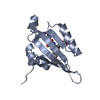

 PDBj
PDBj


 Assembly
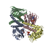

 Mass: 505.208 Da / Num. of mol.: 4 / Source method: obtained synthetically / Formula: C11H18N5O12P3 / Comment: AMP-PCP, energy-carrying molecule analogue*YM
 Mass: 24.305 Da / Num. of mol.: 2 / Source method: obtained synthetically / Formula: Mg
 Sample preparation
 / Beamline: PROXIMA 1 / Wavelength: 0.98011,1.80000
 Processing