 Movie
Movie Controller

[English] 日本語
 Yorodumi
- PDB-2qjp: Crystal structure of wild type rhodobacter sphaeroides with stigm... -
+ Open data

- Basic information
| Entry | Database: PDB / ID: 2qjp | ||||||
|---|---|---|---|---|---|---|---|
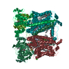
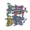
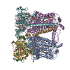
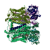
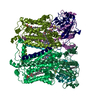
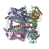
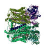
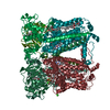
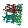
| Title | Crystal structure of wild type rhodobacter sphaeroides with stigmatellin and antimycin inhibited | ||||||
 Components Components |
| ||||||
 Keywords Keywords | ELECTRON TRANSPORT / Cytochrome b with 8 TM helices / one c-term TM in cytochrome c1 and an N-term TM in the iron-sulfur-protein (Rieske) / Heme / Membrane / Metal-binding / Respiratory chain / Transmembrane / Transport / 2Fe-2S / Inner membrane / Oxidoreductase | ||||||
| Function / homology |  Function and homology information Function and homology informationrespiratory chain complex III / quinol-cytochrome-c reductase / quinol-cytochrome-c reductase activity / respiratory electron transport chain / 2 iron, 2 sulfur cluster binding / electron transfer activity / heme binding / metal ion binding / membrane / plasma membrane Similarity search - Function | ||||||
| Biological species |  Rhodobacter sphaeroides (bacteria) Rhodobacter sphaeroides (bacteria) | ||||||
| Method |  X-RAY DIFFRACTION / SYNCHROTRON / MOLECULAR REPLACEMENT / Resolution: 2.6 Å X-RAY DIFFRACTION / SYNCHROTRON / MOLECULAR REPLACEMENT / Resolution: 2.6 Å | ||||||
 Authors Authors | Esser, L. / Xia, D. | ||||||
 Citation Citation | Journal: J.Biol.Chem. / Year: 2008 Title: Inhibitor-complexed structures of the cytochrome bc1 from the photosynthetic bacterium Rhodobacter sphaeroides. Authors: Esser, L. / Elberry, M. / Zhou, F. / Yu, C.A. / Yu, L. / Xia, D. | ||||||
| History |
|
- Structure visualization
| Structure viewer | Molecule: MolmilJmol/JSmol |
|---|
- Downloads & links
-Download
| PDBx/mmCIF format | 2qjp.cif.gz | 703.5 KB | Display | PDBx/mmCIF format |
|---|---|---|---|---|
| PDB format | pdb2qjp.ent.gz | 571.4 KB | Display | PDB format |
| PDBx/mmJSON format | 2qjp.json.gz | Tree view | PDBx/mmJSON format | |
| Others |  Other downloads Other downloads |
-Validation report
| Summary document | 2qjp_validation.pdf.gz | 7.3 MB | Display | wwPDB validaton report |
|---|---|---|---|---|
| Full document | 2qjp_full_validation.pdf.gz | 7.5 MB | Display | |
| Data in XML | 2qjp_validation.xml.gz | 142.4 KB | Display | |
| Data in CIF | 2qjp_validation.cif.gz | 183.1 KB | Display | |
| Arichive directory | https://data.pdbj.org/pub/pdb/validation_reports/qj/2qjpftp://data.pdbj.org/pub/pdb/validation_reports/qj/2qjp | HTTPS FTP |
-Related structure data
| Related structure data | 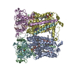 2qjkC 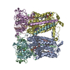 2qjyC 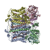 2fynS C: citing same article ( S: Starting model for refinement |
|---|---|
| Similar structure data |


-Links
 PDBj
PDBj


- Assembly
| Deposited unit | 
| ||||||||
|---|---|---|---|---|---|---|---|---|---|
| 1 | 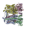
| ||||||||
| 2 | 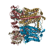
| ||||||||
| Unit cell |
|
-Components
-Protein , 3 types, 12 molecules ADGJBEHKCFIL
| #1: Protein | Mass: 48367.523 Da / Num. of mol.: 4 Source method: isolated from a genetically manipulated source Source: (gene. exp.) Rhodobacter sphaeroides (bacteria) / Gene: petB, fbcB / Plasmid: pRKDfbcFBCQ / Production host: #2: Protein | Mass: 27786.162 Da / Num. of mol.: 4 Source method: isolated from a genetically manipulated source Source: (gene. exp.) Rhodobacter sphaeroides (bacteria) / Gene: fbcC / Plasmid: pRKDfbcFBCQ / Production host: #3: Protein | Mass: 19071.494 Da / Num. of mol.: 4 Source method: isolated from a genetically manipulated source Source: (gene. exp.) Rhodobacter sphaeroides (bacteria) / Gene: petA, fbcF / Plasmid: pRKDfbcFBCQ / Production host: |
|---|
-Sugars , 1 types, 4 molecules 
| #4: Sugar | ChemComp-BGL /  Type: D-saccharide, beta linking / Mass: 292.369 Da / Num. of mol.: 4 Type: D-saccharide, beta linking / Mass: 292.369 Da / Num. of mol.: 4Source method: isolated from a genetically manipulated source Formula: C14H28O6 / Comment: detergent*YM |
|---|
-Non-polymers , 7 types, 243 molecules 


| #5: Chemical | ChemComp-HEM /  Mass: 616.487 Da / Num. of mol.: 12 / Source method: obtained synthetically / Formula: C34H32FeN4O4 Mass: 616.487 Da / Num. of mol.: 12 / Source method: obtained synthetically / Formula: C34H32FeN4O4#6: Chemical | ChemComp-SMA /  Mass: 514.650 Da / Num. of mol.: 4 / Source method: obtained synthetically / Formula: C30H42O7 Mass: 514.650 Da / Num. of mol.: 4 / Source method: obtained synthetically / Formula: C30H42O7#7: Chemical | ChemComp-LOP / (  Mass: 661.890 Da / Num. of mol.: 4 / Source method: obtained synthetically / Formula: C35H68NO8P / Comment: phospholipid*YM Mass: 661.890 Da / Num. of mol.: 4 / Source method: obtained synthetically / Formula: C35H68NO8P / Comment: phospholipid*YM#8: Chemical | ChemComp-ANJ / (  Mass: 548.625 Da / Num. of mol.: 4 / Source method: obtained synthetically / Formula: C28H40N2O9 Mass: 548.625 Da / Num. of mol.: 4 / Source method: obtained synthetically / Formula: C28H40N2O9#9: Chemical | ChemComp-SR /  Mass: 87.620 Da / Num. of mol.: 6 / Source method: obtained synthetically / Formula: Sr Mass: 87.620 Da / Num. of mol.: 6 / Source method: obtained synthetically / Formula: Sr#10: Chemical | ChemComp-FES /  Mass: 175.820 Da / Num. of mol.: 4 / Source method: obtained synthetically / Formula: Fe2S2 Mass: 175.820 Da / Num. of mol.: 4 / Source method: obtained synthetically / Formula: Fe2S2#11: Water | ChemComp-HOH / | Mass: 18.015 Da / Num. of mol.: 209 / Source method: isolated from a natural source / Formula: H2O |
|---|
-Details
| Has protein modification | Y |
|---|
-Experimental details
-Experiment
| Experiment | Method: X-RAY DIFFRACTION / Number of used crystals: 1 |
|---|
- Sample preparation
| Crystal | Density Matthews: 3.44 Å3/Da / Density % sol: 64.22 % |
|---|---|
| Crystal grow | Temperature: 288 K / Method: vapor diffusion, sitting drop / pH: 7.5 Details: 50 mM Tris, 0.5% b-OG, 0.12% Sucrosemonocaprate, 6.39% PEG400, 1.50% PEG3350, 10 mM Strontium nitrate, pH 7.5, VAPOR DIFFUSION, SITTING DROP, temperature 288K |
-Data collection
| Diffraction | Mean temperature: 100 K |
|---|---|
| Diffraction source | Source: SYNCHROTRON / Site: APS  / Beamline: 22-ID / Wavelength: 0.75 Å / Beamline: 22-ID / Wavelength: 0.75 Å |
| Detector | Type: MARRESEARCH / Detector: CCD / Date: Aug 11, 2006 / Details: mirrors |
| Radiation | Monochromator: Silicon 111 / Protocol: SINGLE WAVELENGTH / Monochromatic (M) / Laue (L): M / Scattering type: x-ray |
| Radiation wavelength | Wavelength: 0.75 Å / Relative weight: 1 |
| Reflection | Resolution: 2.6→50 Å / Num. all: 157560 / Num. obs: 156392 / % possible obs: 98.8 % / Observed criterion σ(F): 0 / Observed criterion σ(I): -1.5 / Redundancy: 4.5 % / Biso Wilson estimate: 54.3 Å2 / Rmerge(I) obs: 0.11 / Rsym value: 0.11 / Net I/σ(I): 10.51 |
| Reflection shell | Resolution: 2.6→2.69 Å / Redundancy: 2.8 % / Rmerge(I) obs: 0.67 / Mean I/σ(I) obs: 1.42 / Num. unique all: 14848 / Rsym value: 0.67 / % possible all: 94.3 |
- Processing
| Software |
| ||||||||||||||||||||||||||||||||||||
|---|---|---|---|---|---|---|---|---|---|---|---|---|---|---|---|---|---|---|---|---|---|---|---|---|---|---|---|---|---|---|---|---|---|---|---|---|---|
| Refinement | Method to determine structure: MOLECULAR REPLACEMENT Starting model: pdb entry 2FYN Resolution: 2.6→17.98 Å / Rfactor Rfree error: 0.005 / Data cutoff high absF: 3101164.84 / Data cutoff low absF: 0 / Isotropic thermal model: RESTRAINED / Cross valid method: THROUGHOUT / σ(F): 0 / Stereochemistry target values: Engh & Huber / Details: NCS restraints
| ||||||||||||||||||||||||||||||||||||
| Solvent computation | Solvent model: FLAT MODEL / Bsol: 56.4758 Å2 / ksol: 0.335398 e/Å3 | ||||||||||||||||||||||||||||||||||||
| Displacement parameters | Biso mean: 78.5 Å2
| ||||||||||||||||||||||||||||||||||||
| Refine analyze |
| ||||||||||||||||||||||||||||||||||||
| Refinement step | Cycle: LAST / Resolution: 2.6→17.98 Å
| ||||||||||||||||||||||||||||||||||||
| Refine LS restraints |
| ||||||||||||||||||||||||||||||||||||
| Refine LS restraints NCS | NCS model details: CONSTR | ||||||||||||||||||||||||||||||||||||
| LS refinement shell | Resolution: 2.6→2.76 Å / Rfactor Rfree error: 0.02 / Total num. of bins used: 6
| ||||||||||||||||||||||||||||||||||||
| Xplor file |
|
