 Movie
Movie Controller


+ Open data

- Basic information
| Entry | Database: PDB / ID: 1kb9 | ||||||
|---|---|---|---|---|---|---|---|
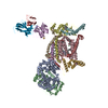
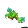
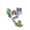
| Title | YEAST CYTOCHROME BC1 COMPLEX | ||||||
 Components Components |
| ||||||
 Keywords Keywords | OXIDOREDUCTASE/ELECTRON TRANSPORT / oxidoreductase / ubiquinone / stigmatellin / cardiolipin / phosphatidylinositol / phosphatidylcholin / phosphatidylethanolamin / undecyl-maltopyranoside / OXIDOREDUCTASE-ELECTRON TRANSPORT COMPLEX | ||||||
| Function / homology |  Function and homology information Function and homology information: / Complex III assembly / matrix side of mitochondrial inner membrane / Respiratory electron transport / : / mitochondrial processing peptidase complex / Mitochondrial protein degradation / mitochondrial respiratory chain complex III assembly / respiratory chain complex III / cellular respiration ...: / Complex III assembly / matrix side of mitochondrial inner membrane / Respiratory electron transport / : / mitochondrial processing peptidase complex / Mitochondrial protein degradation / mitochondrial respiratory chain complex III assembly / respiratory chain complex III / cellular respiration / quinol-cytochrome-c reductase / quinol-cytochrome-c reductase activity / mitochondrial electron transport, ubiquinol to cytochrome c / mitochondrial crista / proton transmembrane transport / nuclear periphery / aerobic respiration / metalloendopeptidase activity / mitochondrial intermembrane space / 2 iron, 2 sulfur cluster binding / oxidoreductase activity / mitochondrial inner membrane / heme binding / mitochondrion / proteolysis / membrane / metal ion binding / cytosol Similarity search - Function | ||||||
| Biological species |   | ||||||
| Method |  X-RAY DIFFRACTION / Resolution: 2.3 Å X-RAY DIFFRACTION / Resolution: 2.3 Å | ||||||
 Authors Authors | Lange, C. / Nett, J.H. / Trumpower, B.L. / Hunte, C. | ||||||
 Citation Citation | Journal: EMBO J. / Year: 2001 Title: SPECIFIC ROLES OF PROTEIN-PHOSPHOLIPID INTERACTIONS IN THE YEAST CYTOCHROME BC1 COMPLEX STRUCTURE Authors: Lange, C. / Nett, J.H. / Trumpower, B.L. / Hunte, C. #1: Journal: Structure / Year: 2000Title: Structure Of The Yeast Cytochrome Bc1 Complex Co-Crystallized With An Antibody Fv-Fragment Authors: Hunte, C. / Koepke, J. / Lange, C. / Rossmanith, T. / Michel, H. | ||||||
| History |
|
- Structure visualization
| Structure viewer | Molecule: MolmilJmol/JSmol |
|---|
- Downloads & links
-Download
| PDBx/mmCIF format | 1kb9.cif.gz | 477.8 KB | Display | PDBx/mmCIF format |
|---|---|---|---|---|
| PDB format | pdb1kb9.ent.gz | 371.1 KB | Display | PDB format |
| PDBx/mmJSON format | 1kb9.json.gz | Tree view | PDBx/mmJSON format | |
| Others |  Other downloads Other downloads |
-Validation report
| Arichive directory | https://data.pdbj.org/pub/pdb/validation_reports/kb/1kb9ftp://data.pdbj.org/pub/pdb/validation_reports/kb/1kb9 | HTTPS FTP |
|---|
-Related structure data
| Related structure data | |
|---|---|
| Similar structure data |


-Links
 PDBj
PDBj


- Assembly
| Deposited unit | 
| ||||||||
|---|---|---|---|---|---|---|---|---|---|
| 1 |
| ||||||||
| Unit cell |
|
-Components
-UBIQUINOL-CYTOCHROME C REDUCTASE COMPLEX ... , 6 types, 6 molecules ABFGHI
| #1: Protein | Mass: 47445.242 Da / Num. of mol.: 1 / Fragment: RESIDUES 27-457 Source method: isolated from a genetically manipulated source Source: (gene. exp.) Organelle: MITOCHONDRIA / References: UniProt: P07256, quinol-cytochrome-c reductase |
|---|---|
| #2: Protein | Mass: 38751.918 Da / Num. of mol.: 1 / Fragment: RESIDUES 17-368 Source method: isolated from a genetically manipulated source Source: (gene. exp.) Organelle: MITOCHONDRIA / References: UniProt: P07257, quinol-cytochrome-c reductase |
| #6: Protein | Mass: 8854.792 Da / Num. of mol.: 1 / Fragment: RESIDUES 74-147 Source method: isolated from a genetically manipulated source Source: (gene. exp.) Organelle: MITOCHONDRIA / References: UniProt: P00127, quinol-cytochrome-c reductase |
| #7: Protein | Mass: 14355.443 Da / Num. of mol.: 1 / Fragment: RESIDUES 3-127 Source method: isolated from a genetically manipulated source Source: (gene. exp.) Organelle: MITOCHONDRIA / References: UniProt: P00128, quinol-cytochrome-c reductase |
| #8: Protein | Mass: 10856.314 Da / Num. of mol.: 1 / Fragment: RESIDUES 2-94 Source method: isolated from a genetically manipulated source Source: (gene. exp.) Organelle: MITOCHONDRIA / References: UniProt: P08525, quinol-cytochrome-c reductase |
| #9: Protein | Mass: 6301.232 Da / Num. of mol.: 1 / Fragment: RESIDUES 4-58 Source method: isolated from a genetically manipulated source Source: (gene. exp.) Organelle: MITOCHONDRIA / References: UniProt: P22289, quinol-cytochrome-c reductase |
-Protein , 3 types, 3 molecules CDE
| #3: Protein | Mass: 43674.535 Da / Num. of mol.: 1 Source method: isolated from a genetically manipulated source Source: (gene. exp.) Organelle: MITOCHONDRIA / References: UniProt: P00163 |
|---|---|
| #4: Protein | Mass: 27521.020 Da / Num. of mol.: 1 / Fragment: RESIDUES 62-307 Source method: isolated from a genetically manipulated source Source: (gene. exp.) Organelle: MITOCHONDRIA / References: UniProt: P07143 |
| #5: Protein | Mass: 20122.955 Da / Num. of mol.: 1 / Fragment: RESIDUES 31-215 Source method: isolated from a genetically manipulated source Source: (gene. exp.) Organelle: MITOCHONDRIA / References: UniProt: P08067, quinol-cytochrome-c reductase |
-Antibody , 2 types, 2 molecules JK
| #10: Antibody | Mass: 14365.817 Da / Num. of mol.: 1 Source method: isolated from a genetically manipulated source Source: (gene. exp.)  |
|---|---|
| #11: Antibody | Mass: 11926.350 Da / Num. of mol.: 1 Source method: isolated from a genetically manipulated source Source: (gene. exp.) |
-Non-polymers , 10 types, 333 molecules 


| #12: Chemical | ChemComp-PCF /  Mass: 734.039 Da / Num. of mol.: 1 / Source method: obtained synthetically / Formula: C40H80NO8P Mass: 734.039 Da / Num. of mol.: 1 / Source method: obtained synthetically / Formula: C40H80NO8P | ||||||||||||||
|---|---|---|---|---|---|---|---|---|---|---|---|---|---|---|---|
| #13: Chemical | ChemComp-UMQ /  Mass: 496.589 Da / Num. of mol.: 1 / Source method: obtained synthetically / Formula: C23H44O11 / Comment: detergent*YM Mass: 496.589 Da / Num. of mol.: 1 / Source method: obtained synthetically / Formula: C23H44O11 / Comment: detergent*YM | ||||||||||||||
| #14: Chemical |  Mass: 616.487 Da / Num. of mol.: 3 / Source method: obtained synthetically / Formula: C34H32FeN4O4 Mass: 616.487 Da / Num. of mol.: 3 / Source method: obtained synthetically / Formula: C34H32FeN4O4#15: Chemical | ChemComp-SMA / |  Mass: 514.650 Da / Num. of mol.: 1 / Source method: obtained synthetically / Formula: C30H42O7 Mass: 514.650 Da / Num. of mol.: 1 / Source method: obtained synthetically / Formula: C30H42O7#16: Chemical | ChemComp-UQ6 / |  Mass: 592.891 Da / Num. of mol.: 1 / Source method: obtained synthetically / Formula: C39H60O4 Mass: 592.891 Da / Num. of mol.: 1 / Source method: obtained synthetically / Formula: C39H60O4#17: Chemical | ChemComp-PIE / |  Mass: 836.061 Da / Num. of mol.: 1 / Source method: obtained synthetically / Formula: C43H80O13P Mass: 836.061 Da / Num. of mol.: 1 / Source method: obtained synthetically / Formula: C43H80O13P#18: Chemical |  Mass: 691.959 Da / Num. of mol.: 2 / Source method: obtained synthetically / Formula: C37H74NO8P / Comment: phospholipid*YM Mass: 691.959 Da / Num. of mol.: 2 / Source method: obtained synthetically / Formula: C37H74NO8P / Comment: phospholipid*YM#19: Chemical | ChemComp-CDL / |  Mass: 1464.043 Da / Num. of mol.: 1 / Source method: obtained synthetically / Formula: C81H156O17P2 / Comment: phospholipid*YM Mass: 1464.043 Da / Num. of mol.: 1 / Source method: obtained synthetically / Formula: C81H156O17P2 / Comment: phospholipid*YM#20: Chemical | ChemComp-FES / |  Mass: 175.820 Da / Num. of mol.: 1 / Source method: obtained synthetically / Formula: Fe2S2 Mass: 175.820 Da / Num. of mol.: 1 / Source method: obtained synthetically / Formula: Fe2S2#21: Water | ChemComp-HOH / | Mass: 18.015 Da / Num. of mol.: 321 / Source method: isolated from a natural source / Formula: H2O |
-Details
| Has protein modification | Y |
|---|
-Experimental details
-Experiment
| Experiment | Method: X-RAY DIFFRACTION / Number of used crystals: 10 |
|---|
- Sample preparation
| Crystal | Density Matthews: 4.7 Å3/Da / Density % sol: 73.83 % |
|---|---|
| Crystal grow | Method: vapor diffusion, sitting drop / pH: 8 / Details: PEG4000, pH 8, VAPOR DIFFUSION, SITTING DROP |
| Crystal grow | *PLUS Method: unknown / Details: Hunte, C., (2000) Structure, 8, 669. |
-Data collection
| Detector | Type: MARRESEARCH / Detector: CCD |
|---|---|
| Radiation | Protocol: SINGLE WAVELENGTH / Monochromatic (M) / Laue (L): M / Scattering type: x-ray |
| Radiation wavelength | Relative weight: 1 |
| Reflection | Resolution: 2.3→14.96 Å / Num. all: 168517 / Num. obs: 168517 / Observed criterion σ(F): 0 / Biso Wilson estimate: 31.9 Å2 / Limit h max: 82 / Limit h min: -93 / Limit k max: 71 / Limit k min: -93 / Limit l max: 63 / Limit l min: 0 / Observed criterion F max: 3621196.32 / Observed criterion F min: 18.17 |
| Reflection | *PLUS Highest resolution: 2.3 Å |
- Processing
| Software |
| ||||||||||||||||||||||||||||||||||||||||||||||||||||||||||||||||||||||||||||||||||||||||||
|---|---|---|---|---|---|---|---|---|---|---|---|---|---|---|---|---|---|---|---|---|---|---|---|---|---|---|---|---|---|---|---|---|---|---|---|---|---|---|---|---|---|---|---|---|---|---|---|---|---|---|---|---|---|---|---|---|---|---|---|---|---|---|---|---|---|---|---|---|---|---|---|---|---|---|---|---|---|---|---|---|---|---|---|---|---|---|---|---|---|---|---|
| Refinement | Resolution: 2.3→14.96 Å / Rfactor Rfree error: 0.004 / Occupancy max: 1 / Occupancy min: 0 / Cross valid method: THROUGHOUT
| ||||||||||||||||||||||||||||||||||||||||||||||||||||||||||||||||||||||||||||||||||||||||||
| Solvent computation | Solvent model: CNS bulk solvent model used / Bsol: 38.6265 Å2 / ksol: 0.273859 e/Å3 | ||||||||||||||||||||||||||||||||||||||||||||||||||||||||||||||||||||||||||||||||||||||||||
| Displacement parameters | Biso max: 157.38 Å2 / Biso mean: 69.92 Å2 / Biso min: 20.92 Å2
| ||||||||||||||||||||||||||||||||||||||||||||||||||||||||||||||||||||||||||||||||||||||||||
| Refine analyze |
| ||||||||||||||||||||||||||||||||||||||||||||||||||||||||||||||||||||||||||||||||||||||||||
| Refinement step | Cycle: LAST / Resolution: 2.3→14.96 Å
| ||||||||||||||||||||||||||||||||||||||||||||||||||||||||||||||||||||||||||||||||||||||||||
| Refine LS restraints |
| ||||||||||||||||||||||||||||||||||||||||||||||||||||||||||||||||||||||||||||||||||||||||||
| LS refinement shell | Refine-ID: X-RAY DIFFRACTION / Total num. of bins used: 8
| ||||||||||||||||||||||||||||||||||||||||||||||||||||||||||||||||||||||||||||||||||||||||||
| Xplor file |
| ||||||||||||||||||||||||||||||||||||||||||||||||||||||||||||||||||||||||||||||||||||||||||
| Software | *PLUS Name: CNS / Classification: refinement | ||||||||||||||||||||||||||||||||||||||||||||||||||||||||||||||||||||||||||||||||||||||||||
| Refinement | *PLUS Highest resolution: 2.3 Å / Lowest resolution: 15 Å | ||||||||||||||||||||||||||||||||||||||||||||||||||||||||||||||||||||||||||||||||||||||||||
| Solvent computation | *PLUS | ||||||||||||||||||||||||||||||||||||||||||||||||||||||||||||||||||||||||||||||||||||||||||
| Displacement parameters | *PLUS | ||||||||||||||||||||||||||||||||||||||||||||||||||||||||||||||||||||||||||||||||||||||||||
| Refine LS restraints | *PLUS
|
