 Movie
Movie Controller

[English] 日本語
 Yorodumi
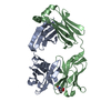
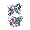
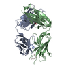
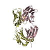
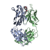
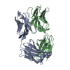
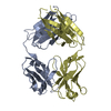
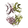
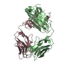
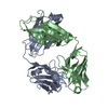
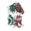
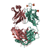
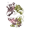
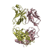
- PDB-2pr4: Crystal Structure of Fab' from the HIV-1 Neutralizing Antibody 2F5 -
+ Open data

- Basic information
| Entry | Database: PDB / ID: 2pr4 | |||||||||
|---|---|---|---|---|---|---|---|---|---|---|
| Title | Crystal Structure of Fab' from the HIV-1 Neutralizing Antibody 2F5 | |||||||||
 Components Components |
| |||||||||
 Keywords Keywords | VIRAL PROTEIN / nmAb 2F5 / gp41 / HIV | |||||||||
| Function / homology | Immunoglobulins / Immunoglobulin-like / Sandwich / Mainly Beta Function and homology information Function and homology information | |||||||||
| Biological species |  Homo sapiens (human) Homo sapiens (human) | |||||||||
| Method |  X-RAY DIFFRACTION / MOLECULAR REPLACEMENT / Resolution: 2.05 Å X-RAY DIFFRACTION / MOLECULAR REPLACEMENT / Resolution: 2.05 Å | |||||||||
 Authors Authors | Bryson, S. / Julien, J.-P. / Pai, E.F. | |||||||||
 Citation Citation | Journal: J.Mol.Biol. / Year: 2008 Title: Structural details of HIV-1 recognition by the broadly neutralizing monoclonal antibody 2F5: epitope conformation, antigen-recognition loop mobility, and anion-binding site. Authors: Julien, J.P. / Bryson, S. / Nieva, J.L. / Pai, E.F. | |||||||||
| History |
|
- Structure visualization
| Structure viewer | Molecule: MolmilJmol/JSmol |
|---|
- Downloads & links
-Download
| PDBx/mmCIF format | 2pr4.cif.gz | 98.7 KB | Display | PDBx/mmCIF format |
|---|---|---|---|---|
| PDB format | pdb2pr4.ent.gz | 74.6 KB | Display | PDB format |
| PDBx/mmJSON format | 2pr4.json.gz | Tree view | PDBx/mmJSON format | |
| Others |  Other downloads Other downloads |
-Validation report
| Arichive directory | https://data.pdbj.org/pub/pdb/validation_reports/pr/2pr4ftp://data.pdbj.org/pub/pdb/validation_reports/pr/2pr4 | HTTPS FTP |
|---|
-Related structure data
| Related structure data | 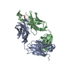 2p8lC 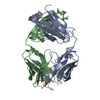 2p8mC 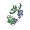 2p8pC 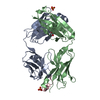 3d0lC  3d0vC 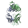 3droC 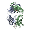 3drqC C: citing same article ( |
|---|---|
| Similar structure data |


-Links
 PDBj
PDBj


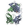
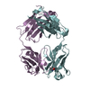
- Assembly
| Deposited unit | 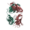
| ||||||||
|---|---|---|---|---|---|---|---|---|---|
| 1 |
| ||||||||
| Unit cell |
| ||||||||
| Details | Light chain interacts with the Heavy chain to form Fab' fragment. |
-Components
| #1: Antibody | Mass: 23363.844 Da / Num. of mol.: 1 / Source method: isolated from a natural source / Source: (natural) Homo sapiens (human) |
|---|---|
| #2: Antibody | Mass: 25001.436 Da / Num. of mol.: 1 / Source method: isolated from a natural source / Source: (natural) Homo sapiens (human) |
| #3: Water | ChemComp-HOH /  Mass: 18.015 Da / Num. of mol.: 266 / Source method: isolated from a natural source / Formula: H2O Mass: 18.015 Da / Num. of mol.: 266 / Source method: isolated from a natural source / Formula: H2O |
| Has protein modification | Y |
-Experimental details
-Experiment
| Experiment | Method: X-RAY DIFFRACTION / Number of used crystals: 1 |
|---|
- Sample preparation
| Crystal | Density Matthews: 2.38 Å3/Da / Density % sol: 48.27 % |
|---|---|
| Crystal grow | Temperature: 293 K / Method: vapor diffusion, hanging drop / pH: 6.5 Details: 0.5M Li2SO4, 15% PEG 8000, pH 6.5, VAPOR DIFFUSION, HANGING DROP, temperature 293K |
-Data collection
| Diffraction | Mean temperature: 105 K |
|---|---|
| Diffraction source | Source: ROTATING ANODE / Type: RIGAKU / Wavelength: 1.5418 Å |
| Detector | Type: SIEMENS / Detector: AREA DETECTOR / Date: Oct 1, 1998 |
| Radiation | Monochromator: NI FILTER / Protocol: SINGLE WAVELENGTH / Monochromatic (M) / Laue (L): M / Scattering type: x-ray |
| Radiation wavelength | Wavelength: 1.5418 Å / Relative weight: 1 |
| Reflection | Resolution: 2.05→20 Å / Num. obs: 26917 / % possible obs: 92 % / Redundancy: 3.2 % / Biso Wilson estimate: 11.7 Å2 / Net I/σ(I): 15 |
| Reflection shell | Resolution: 2.05→2.1 Å / Redundancy: 3 % / Mean I/σ(I) obs: 3.8 / Rsym value: 0.313 / % possible all: 93 |
- Processing
| Software |
| ||||||||||||||||
|---|---|---|---|---|---|---|---|---|---|---|---|---|---|---|---|---|---|
| Refinement | Method to determine structure: MOLECULAR REPLACEMENT / Resolution: 2.05→20 Å / σ(I): 0
| ||||||||||||||||
| Refine analyze |
| ||||||||||||||||
| Refinement step | Cycle: LAST / Resolution: 2.05→20 Å
| ||||||||||||||||
| Refine LS restraints |
| ||||||||||||||||
| LS refinement shell | Resolution: 2.05→2.18 Å
|
