 Movie
Movie Controller

[English] 日本語
 Yorodumi
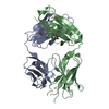
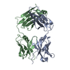
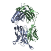
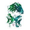
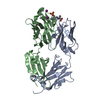
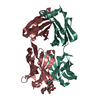
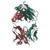
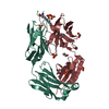
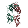
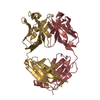
- PDB-3drq: Crystal structure of the HIV-1 broadly neutralizing antibody 2F5 ... -
+ Open data

- Basic information
| Entry | Database: PDB / ID: 3drq | ||||||
|---|---|---|---|---|---|---|---|
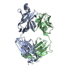
| Title | Crystal structure of the HIV-1 broadly neutralizing antibody 2F5 in complex with the gp41 FP-MPER Hyb3K construct 514GIGALFLGFLGAAGS528KK-Ahx-655KNEQELLELDKWASLWN671 soaked in PEG/2-propanol solution | ||||||
 Components Components |
| ||||||
 Keywords Keywords | IMMUNE SYSTEM / HIV-1 / 2F5 / gp41 / nmAb | ||||||
| Function / homology |  Function and homology information Function and homology informationimmunoglobulin complex / adaptive immune response / extracellular region / plasma membrane Similarity search - Function | ||||||
| Biological species |  Homo sapiens (human) Homo sapiens (human) | ||||||
| Method |  X-RAY DIFFRACTION / MOLECULAR REPLACEMENT / Resolution: 2 Å X-RAY DIFFRACTION / MOLECULAR REPLACEMENT / Resolution: 2 Å | ||||||
 Authors Authors | Bryson, S. / Julien, J.P. / Pai, E.F. | ||||||
 Citation Citation | Journal: J.Mol.Biol. / Year: 2008 Title: Structural details of HIV-1 recognition by the broadly neutralizing monoclonal antibody 2F5: epitope conformation, antigen-recognition loop mobility, and anion-binding site. Authors: Julien, J.P. / Bryson, S. / Nieva, J.L. / Pai, E.F. | ||||||
| History |
|
- Structure visualization
| Structure viewer | Molecule: MolmilJmol/JSmol |
|---|
- Downloads & links
-Download
| PDBx/mmCIF format | 3drq.cif.gz | 103 KB | Display | PDBx/mmCIF format |
|---|---|---|---|---|
| PDB format | pdb3drq.ent.gz | 76.7 KB | Display | PDB format |
| PDBx/mmJSON format | 3drq.json.gz | Tree view | PDBx/mmJSON format | |
| Others |  Other downloads Other downloads |
-Validation report
| Arichive directory | https://data.pdbj.org/pub/pdb/validation_reports/dr/3drqftp://data.pdbj.org/pub/pdb/validation_reports/dr/3drq | HTTPS FTP |
|---|
-Related structure data
| Related structure data | 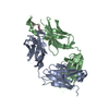 2p8lC 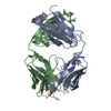 2p8mC 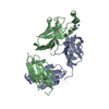 2p8pC 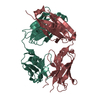 2pr4C 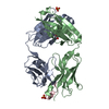 3d0lSC  3d0vC 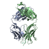 3droC  3drv S: Starting model for refinement C: citing same article ( |
|---|---|
| Similar structure data |


-Links
 PDBj
PDBj


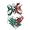
- Assembly
| Deposited unit | 
| ||||||||
|---|---|---|---|---|---|---|---|---|---|
| 1 |
| ||||||||
| Unit cell |
|
-Components
| #1: Antibody | Mass: 23363.844 Da / Num. of mol.: 1 Source method: isolated from a genetically manipulated source Source: (gene. exp.) Homo sapiens (human) / Cell line (production host): hybridoma cells |
|---|---|
| #2: Antibody | Mass: 24985.436 Da / Num. of mol.: 1 Source method: isolated from a genetically manipulated source Source: (gene. exp.) Homo sapiens (human) / Cell line (production host): hybridoma cells / References: UniProt: Q6PYX1*PLUS |
| #3: Protein/peptide | Mass: 3822.411 Da / Num. of mol.: 1 / Source method: obtained synthetically Details: The peptide was chemically synthesized by Fmoc chemistry. The sequence of the peptide is naturally found in the human immunodeficiency virus. |
| #4: Chemical | ChemComp-GOL /   Mass: 92.094 Da / Num. of mol.: 1 / Source method: obtained synthetically / Formula: C3H8O3 Mass: 92.094 Da / Num. of mol.: 1 / Source method: obtained synthetically / Formula: C3H8O3 |
| #5: Water | ChemComp-HOH /  Mass: 18.015 Da / Num. of mol.: 234 / Source method: isolated from a natural source / Formula: H2O Mass: 18.015 Da / Num. of mol.: 234 / Source method: isolated from a natural source / Formula: H2O |
| Has protein modification | Y |
| Nonpolymer details | ACA IN THE PEPTIDE SEQUENCE STANDS FOR 6-AMINO-HEXANOIC ACID LINKER. |
-Experimental details
-Experiment
| Experiment | Method: X-RAY DIFFRACTION / Number of used crystals: 1 |
|---|
- Sample preparation
| Crystal | Density Matthews: 2.2 Å3/Da / Density % sol: 43.98 % |
|---|---|
| Crystal grow | Temperature: 293 K / Method: vapor diffusion, hanging drop / pH: 5.6 Details: Crystals grown in 0.1 M Nacitrate pH 5.6, 1.6 M ammonium sulfate, 0.01% Tween-20 and soaked for 36h in 0.1 M Nacitrate pH 5.6, 16% PEG 4K, 16% 2-propanol, VAPOR DIFFUSION, HANGING DROP, temperature 293K |
-Data collection
| Diffraction | Mean temperature: 100 K |
|---|---|
| Diffraction source | Source: ROTATING ANODE / Type: RIGAKU RU200 / Wavelength: 1.54 Å |
| Detector | Type: RIGAKU RAXIS IV++ / Detector: IMAGE PLATE / Date: Aug 25, 2007 |
| Radiation | Protocol: SINGLE WAVELENGTH / Monochromatic (M) / Laue (L): M / Scattering type: x-ray |
| Radiation wavelength | Wavelength: 1.54 Å / Relative weight: 1 |
| Reflection | Resolution: 2→20 Å / Num. all: 31694 / Num. obs: 31016 / % possible obs: 97.9 % / Redundancy: 3.5 % / Biso Wilson estimate: 16.2 Å2 / Rmerge(I) obs: 0.098 |
| Reflection shell | Resolution: 1.99→2.11 Å / Redundancy: 1.7 % / Rmerge(I) obs: 0.454 / % possible all: 96.1 |
- Processing
| Software |
| ||||||||||||||||||||||||||||||||||||
|---|---|---|---|---|---|---|---|---|---|---|---|---|---|---|---|---|---|---|---|---|---|---|---|---|---|---|---|---|---|---|---|---|---|---|---|---|---|
| Refinement | Method to determine structure: MOLECULAR REPLACEMENT Starting model: 3D0L Resolution: 2→19.9 Å / Rfactor Rfree error: 0.006 / Data cutoff high absF: 1048958.42 / Data cutoff low absF: 0 / Isotropic thermal model: RESTRAINED / Cross valid method: THROUGHOUT / σ(F): 0 / Details: BULK SOLVENT MODEL USED
| ||||||||||||||||||||||||||||||||||||
| Solvent computation | Solvent model: FLAT MODEL / Bsol: 53.1513 Å2 / ksol: 0.4 e/Å3 | ||||||||||||||||||||||||||||||||||||
| Displacement parameters | Biso mean: 26.5 Å2
| ||||||||||||||||||||||||||||||||||||
| Refine analyze |
| ||||||||||||||||||||||||||||||||||||
| Refinement step | Cycle: LAST / Resolution: 2→19.9 Å
| ||||||||||||||||||||||||||||||||||||
| Refine LS restraints |
| ||||||||||||||||||||||||||||||||||||
| LS refinement shell | Resolution: 2→2.13 Å / Rfactor Rfree error: 0.022 / Total num. of bins used: 6
| ||||||||||||||||||||||||||||||||||||
| Xplor file |
|
