 Movie
Movie Controller

[English] 日本語
 Yorodumi
- PDB-2on9: Structure of an amyloid forming peptide VQIVYK from the repeat re... -
+ Open data

- Basic information
| Entry | Database: PDB / ID: 2on9 | ||||||
|---|---|---|---|---|---|---|---|
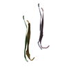
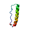
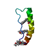
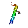
| Title | Structure of an amyloid forming peptide VQIVYK from the repeat region of Tau | ||||||
 Components Components | VQIVYK peptide corresponding to residues 306-311 in the tau protein | ||||||
 Keywords Keywords | PROTEIN FIBRIL / parallel face-to-face-Up/Up beta sheets / steric zipper | ||||||
| Function / homology |  Function and homology information Function and homology informationplus-end-directed organelle transport along microtubule / histone-dependent DNA binding / negative regulation of protein localization to mitochondrion / neurofibrillary tangle / microtubule lateral binding / axonal transport / tubulin complex / positive regulation of protein localization to synapse / phosphatidylinositol bisphosphate binding / generation of neurons ...plus-end-directed organelle transport along microtubule / histone-dependent DNA binding / negative regulation of protein localization to mitochondrion / neurofibrillary tangle / microtubule lateral binding / axonal transport / tubulin complex / positive regulation of protein localization to synapse / phosphatidylinositol bisphosphate binding / generation of neurons / rRNA metabolic process / axonal transport of mitochondrion / axon development / regulation of mitochondrial fission / regulation of microtubule-based movement / central nervous system neuron development / intracellular distribution of mitochondria / regulation of chromosome organization / minor groove of adenine-thymine-rich DNA binding / lipoprotein particle binding / microtubule polymerization / negative regulation of mitochondrial membrane potential / regulation of microtubule polymerization / dynactin binding / apolipoprotein binding / protein polymerization / main axon / Caspase-mediated cleavage of cytoskeletal proteins / regulation of microtubule polymerization or depolymerization / negative regulation of mitochondrial fission / axolemma / glial cell projection / neurofibrillary tangle assembly / positive regulation of axon extension / regulation of cellular response to heat / positive regulation of microtubule polymerization / positive regulation of protein localization / Activation of AMPK downstream of NMDARs / positive regulation of superoxide anion generation / cytoplasmic microtubule organization / cellular response to brain-derived neurotrophic factor stimulus / regulation of calcium-mediated signaling / regulation of long-term synaptic depression / supramolecular fiber organization / axon cytoplasm / somatodendritic compartment / synapse assembly / nuclear periphery / phosphatidylinositol binding / astrocyte activation / protein phosphatase 2A binding / enzyme inhibitor activity / stress granule assembly / regulation of autophagy / regulation of microtubule cytoskeleton organization / cellular response to reactive oxygen species / microglial cell activation / cellular response to nerve growth factor stimulus / Hsp90 protein binding / protein homooligomerization / SH3 domain binding / PKR-mediated signaling / regulation of synaptic plasticity / synapse organization / microtubule cytoskeleton organization / response to lead ion / memory / neuron projection development / cytoplasmic ribonucleoprotein granule / cell-cell signaling / cellular response to heat / single-stranded DNA binding / microtubule cytoskeleton / growth cone / protein-folding chaperone binding / actin binding / double-stranded DNA binding / cell body / microtubule binding / sequence-specific DNA binding / amyloid fibril formation / dendritic spine / microtubule / learning or memory / protein-macromolecule adaptor activity / neuron projection / membrane raft / negative regulation of gene expression / axon / neuronal cell body / DNA damage response / dendrite / protein kinase binding / enzyme binding / mitochondrion / DNA binding / RNA binding / extracellular region / identical protein binding / nucleus Similarity search - Function | ||||||
| Method |  X-RAY DIFFRACTION / SYNCHROTRON / MOLECULAR REPLACEMENT / Resolution: 1.51 Å X-RAY DIFFRACTION / SYNCHROTRON / MOLECULAR REPLACEMENT / Resolution: 1.51 Å | ||||||
 Authors Authors | Sambashivan, S. / Sawaya, M.R. / Eisenberg, D. | ||||||
 Citation Citation | Journal: Nature / Year: 2007 Title: Atomic structures of amyloid cross-beta spines reveal varied steric zippers. Authors: Sawaya, M.R. / Sambashivan, S. / Nelson, R. / Ivanova, M.I. / Sievers, S.A. / Apostol, M.I. / Thompson, M.J. / Balbirnie, M. / Wiltzius, J.J. / McFarlane, H.T. / Madsen, A.O. / Riekel, C. / Eisenberg, D. | ||||||
| History |
|
- Structure visualization
| Structure viewer | Molecule: MolmilJmol/JSmol |
|---|
- Downloads & links
-Download
| PDBx/mmCIF format | 2on9.cif.gz | 10.2 KB | Display | PDBx/mmCIF format |
|---|---|---|---|---|
| PDB format | pdb2on9.ent.gz | 6.4 KB | Display | PDB format |
| PDBx/mmJSON format | 2on9.json.gz | Tree view | PDBx/mmJSON format | |
| Others |  Other downloads Other downloads |
-Validation report
| Arichive directory | https://data.pdbj.org/pub/pdb/validation_reports/on/2on9ftp://data.pdbj.org/pub/pdb/validation_reports/on/2on9 | HTTPS FTP |
|---|
-Related structure data
| Related structure data |  2okzC  2ol9C  2olxC 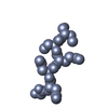 2ommC  2ompC 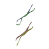 2omqC  2onaC  2onvC  2onwC  2onxC C: citing same article ( |
|---|---|
| Similar structure data |
-Links
 PDBj
PDBj


- Assembly
| Deposited unit | 
| ||||||||
|---|---|---|---|---|---|---|---|---|---|
| 1 | 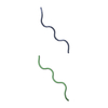
| ||||||||
| Unit cell |
|
-Components
| #1: Protein/peptide | Mass: 749.917 Da / Num. of mol.: 2 / Fragment: residues 306-311 / Source method: obtained synthetically / Details: Peptide was obtained from CSBio company. / References: UniProt: P10636*PLUS #2: Water | ChemComp-HOH / |  Mass: 18.015 Da / Num. of mol.: 7 / Source method: isolated from a natural source / Formula: H2O Mass: 18.015 Da / Num. of mol.: 7 / Source method: isolated from a natural source / Formula: H2O |
|---|
-Experimental details
-Experiment
| Experiment | Method: X-RAY DIFFRACTION / Number of used crystals: 1 |
|---|
- Sample preparation
| Crystal | Density Matthews: 1.53 Å3/Da / Density % sol: 19.71 % |
|---|---|
| Crystal grow | Temperature: 291 K / Method: vapor diffusion, hanging drop / pH: 7.5 Details: Peptide concentration: 30mg/ml, peptide:Reservoir ratio 1:1, Reservoir: 0.2M Ammonium acetate, 0.1M HEPES-Na, 45% MPD, pH 7.5, VAPOR DIFFUSION, HANGING DROP, temperature 291K |
-Data collection
| Diffraction | Mean temperature: 100 K |
|---|---|
| Diffraction source | Source: SYNCHROTRON / Site: ESRF  / Beamline: ID13 / Wavelength: 0.98 / Beamline: ID13 / Wavelength: 0.98 |
| Detector | Type: MAR CCD 165 mm / Detector: CCD / Date: Jul 16, 2005 |
| Radiation | Protocol: SINGLE WAVELENGTH / Monochromatic (M) / Laue (L): M / Scattering type: x-ray |
| Radiation wavelength | Wavelength: 0.98 Å / Relative weight: 1 |
| Reflection | Resolution: 1.5→90 Å / Num. obs: 1222 / % possible obs: 85.4 % / Redundancy: 2.89 % / Rmerge(I) obs: 0.161 / Χ2: 1.058 / Net I/σ(I): 3.8 |
| Reflection shell | Resolution: 1.5→1.62 Å / Rmerge(I) obs: 0.489 / Num. unique all: 238 / Χ2: 1.104 / % possible all: 77 |
- Processing
| Software |
| ||||||||||||||||||||||||||||||||||||||||||||||||||||||||||||||||||||||||||||||||||||||||||||||||||||||||||||||||||||||||
|---|---|---|---|---|---|---|---|---|---|---|---|---|---|---|---|---|---|---|---|---|---|---|---|---|---|---|---|---|---|---|---|---|---|---|---|---|---|---|---|---|---|---|---|---|---|---|---|---|---|---|---|---|---|---|---|---|---|---|---|---|---|---|---|---|---|---|---|---|---|---|---|---|---|---|---|---|---|---|---|---|---|---|---|---|---|---|---|---|---|---|---|---|---|---|---|---|---|---|---|---|---|---|---|---|---|---|---|---|---|---|---|---|---|---|---|---|---|---|---|---|---|
| Refinement | Method to determine structure: MOLECULAR REPLACEMENT Starting model: Extended beta strand VQIVYK Resolution: 1.51→30.96 Å / Cor.coef. Fo:Fc: 0.961 / Cor.coef. Fo:Fc free: 0.964 / SU B: 3.485 / SU ML: 0.113 / Cross valid method: THROUGHOUT / σ(F): 0 / ESU R: 0.139 / ESU R Free: 0.117 / Stereochemistry target values: MAXIMUM LIKELIHOOD / Details: HYDROGENS HAVE BEEN ADDED IN THE RIDING POSITIONS
| ||||||||||||||||||||||||||||||||||||||||||||||||||||||||||||||||||||||||||||||||||||||||||||||||||||||||||||||||||||||||
| Solvent computation | Ion probe radii: 0.8 Å / Shrinkage radii: 0.8 Å / VDW probe radii: 1.4 Å / Solvent model: MASK | ||||||||||||||||||||||||||||||||||||||||||||||||||||||||||||||||||||||||||||||||||||||||||||||||||||||||||||||||||||||||
| Displacement parameters | Biso mean: 8.391 Å2
| ||||||||||||||||||||||||||||||||||||||||||||||||||||||||||||||||||||||||||||||||||||||||||||||||||||||||||||||||||||||||
| Refinement step | Cycle: LAST / Resolution: 1.51→30.96 Å
| ||||||||||||||||||||||||||||||||||||||||||||||||||||||||||||||||||||||||||||||||||||||||||||||||||||||||||||||||||||||||
| Refine LS restraints |
| ||||||||||||||||||||||||||||||||||||||||||||||||||||||||||||||||||||||||||||||||||||||||||||||||||||||||||||||||||||||||
| LS refinement shell | Resolution: 1.51→1.55 Å / Total num. of bins used: 20
|
