 Movie
Movie Controller

[English] 日本語
 Yorodumi
- PDB-2onv: Crystal Structure of the amyloid-fibril forming peptide GGVVIA de... -
+ Open data

- Basic information
| Entry | Database: PDB / ID: 2onv | ||||||
|---|---|---|---|---|---|---|---|
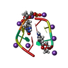
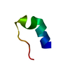
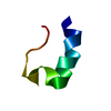
| Title | Crystal Structure of the amyloid-fibril forming peptide GGVVIA derived from the Alzheimer's amyloid Abeta (Abeta37-42). | ||||||
 Components Components | amyloid-fibril forming peptide GGVVIA derived from the Alzheimer's amyloid Abeta | ||||||
 Keywords Keywords | PROTEIN FIBRIL / steric zipper / beta sheets | ||||||
| Method |  X-RAY DIFFRACTION / SYNCHROTRON / MOLECULAR REPLACEMENT / Resolution: 1.61 Å X-RAY DIFFRACTION / SYNCHROTRON / MOLECULAR REPLACEMENT / Resolution: 1.61 Å | ||||||
 Authors Authors | Sambashivan, S. / Sawaya, M.R. / Eisenberg, D. | ||||||
 Citation Citation | Journal: Nature / Year: 2007 Title: Atomic structures of amyloid cross-beta spines reveal varied steric zippers. Authors: Sawaya, M.R. / Sambashivan, S. / Nelson, R. / Ivanova, M.I. / Sievers, S.A. / Apostol, M.I. / Thompson, M.J. / Balbirnie, M. / Wiltzius, J.J. / McFarlane, H.T. / Madsen, A.O. / Riekel, C. / Eisenberg, D. | ||||||
| History |
|
- Structure visualization
| Structure viewer | Molecule:  MolmilJmol/JSmol MolmilJmol/JSmol |
|---|
- Downloads & links
-Download
| PDBx/mmCIF format | 2onv.cif.gz | 7.9 KB | Display | PDBx/mmCIF format |
|---|---|---|---|---|
| PDB format | pdb2onv.ent.gz | 4.9 KB | Display | PDB format |
| PDBx/mmJSON format | 2onv.json.gz | Tree view | PDBx/mmJSON format | |
| Others |  Other downloads Other downloads |
-Validation report
| Arichive directory | https://data.pdbj.org/pub/pdb/validation_reports/on/2onvftp://data.pdbj.org/pub/pdb/validation_reports/on/2onv | HTTPS FTP |
|---|
-Related structure data
| Related structure data |  2okzC  2ol9C  2olxC 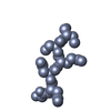 2ommC  2ompC 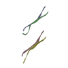 2omqC  2on9C  2onaC  2onwC  2onxC C: citing same article ( |
|---|---|
| Similar structure data |


-Links
 PDBj
PDBj- Assembly
| Deposited unit | 
| ||||||||
|---|---|---|---|---|---|---|---|---|---|
| 1 | 
| ||||||||
| Unit cell |
| ||||||||
| Details | The biological unit is a pair of sheets. Beta strands within the sheet are generated by unit cell translations along the z-axis. The second sheet in the pair of sheets is generated by applying the symmetry operator x+1/2, -y+1/2, -z. Within the second sheet also, beta strands are generated by unit cell translations along the z-axis. |
-Components
| #1: Protein/peptide | Mass: 514.616 Da / Num. of mol.: 1 / Fragment: residues 37-42 / Source method: obtained synthetically / Details: Peptide was commercially synthesized by CSBio. |
|---|---|
| #2: Water | ChemComp-HOH /  Mass: 18.015 Da / Num. of mol.: 3 / Source method: isolated from a natural source / Formula: H2O Mass: 18.015 Da / Num. of mol.: 3 / Source method: isolated from a natural source / Formula: H2O |
-Experimental details
-Experiment
| Experiment | Method: X-RAY DIFFRACTION / Number of used crystals: 1 |
|---|
- Sample preparation
| Crystal | Density Matthews: 1.6 Å3/Da / Density % sol: 23.31 % |
|---|---|
| Crystal grow | Temperature: 291 K / Method: vapor diffusion, hanging drop Details: Peptide concentration: 15.0 mg/ml, Peptide:reservoir:additive ratio 5:4:1, Reservoir: 2.0M Ammonium sulfate, Additive: 3.0% 0.1M hexamine cobalt (III) chloride , VAPOR DIFFUSION, HANGING DROP, temperature 291K |
-Data collection
| Diffraction | Mean temperature: 100 K |
|---|---|
| Diffraction source | Source: SYNCHROTRON / Site: ESRF  / Beamline: ID13 / Wavelength: 0.9466 / Beamline: ID13 / Wavelength: 0.9466 |
| Detector | Type: MAR CCD 165 mm / Detector: CCD / Date: Dec 17, 2005 |
| Radiation | Protocol: SINGLE WAVELENGTH / Monochromatic (M) / Laue (L): M / Scattering type: x-ray |
| Radiation wavelength | Wavelength: 0.9466 Å / Relative weight: 1 |
| Reflection | Resolution: 1.6→90 Å / Num. obs: 532 / % possible obs: 96.7 % / Redundancy: 4.5 % / Biso Wilson estimate: 15.962 Å2 / Rmerge(I) obs: 0.192 / Χ2: 1.085 / Net I/σ(I): 12.6 |
| Reflection shell | Resolution: 1.6→1.72 Å / Redundancy: 4.9 % / Rmerge(I) obs: 0.42 / Num. unique all: 87 / Χ2: 1.102 / % possible all: 96.7 |
- Processing
| Software |
| |||||||||||||||||||||||||||||||||||||||||||||||||||||||||||||||||||||||||||||||||||||||||||||||||||||||||||||||||||
|---|---|---|---|---|---|---|---|---|---|---|---|---|---|---|---|---|---|---|---|---|---|---|---|---|---|---|---|---|---|---|---|---|---|---|---|---|---|---|---|---|---|---|---|---|---|---|---|---|---|---|---|---|---|---|---|---|---|---|---|---|---|---|---|---|---|---|---|---|---|---|---|---|---|---|---|---|---|---|---|---|---|---|---|---|---|---|---|---|---|---|---|---|---|---|---|---|---|---|---|---|---|---|---|---|---|---|---|---|---|---|---|---|---|---|---|---|
| Refinement | Method to determine structure: MOLECULAR REPLACEMENT Starting model: Extended beta strand GGVVIA Resolution: 1.61→20.57 Å / Cor.coef. Fo:Fc: 0.957 / Cor.coef. Fo:Fc free: 0.852 / SU B: 4.479 / SU ML: 0.136 / Cross valid method: THROUGHOUT / σ(F): 0 / ESU R: 0.137 / ESU R Free: 0.146 / Stereochemistry target values: MAXIMUM LIKELIHOOD / Details: HYDROGENS HAVE BEEN ADDED IN THE RIDING POSITIONS
| |||||||||||||||||||||||||||||||||||||||||||||||||||||||||||||||||||||||||||||||||||||||||||||||||||||||||||||||||||
| Solvent computation | Ion probe radii: 0.8 Å / Shrinkage radii: 0.8 Å / VDW probe radii: 1.4 Å / Solvent model: MASK | |||||||||||||||||||||||||||||||||||||||||||||||||||||||||||||||||||||||||||||||||||||||||||||||||||||||||||||||||||
| Displacement parameters | Biso mean: 9.99 Å2
| |||||||||||||||||||||||||||||||||||||||||||||||||||||||||||||||||||||||||||||||||||||||||||||||||||||||||||||||||||
| Refinement step | Cycle: LAST / Resolution: 1.61→20.57 Å /
| |||||||||||||||||||||||||||||||||||||||||||||||||||||||||||||||||||||||||||||||||||||||||||||||||||||||||||||||||||
| Refine LS restraints |
| |||||||||||||||||||||||||||||||||||||||||||||||||||||||||||||||||||||||||||||||||||||||||||||||||||||||||||||||||||
| LS refinement shell | Resolution: 1.61→1.653 Å / Total num. of bins used: 20
|
