 Movie
Movie Controller

[English] 日本語
 Yorodumi
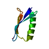
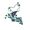
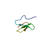
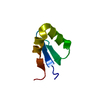
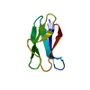
- PDB-2laz: Structure of the first WW domain of human Smurf1 in complex with ... -
+ Open data

- Basic information
| Entry | Database: PDB / ID: 2laz | ||||||
|---|---|---|---|---|---|---|---|
| Title | Structure of the first WW domain of human Smurf1 in complex with a mono-phosphorylated human Smad1 derived peptide | ||||||
 Components Components |
| ||||||
 Keywords Keywords | SIGNALING PROTEIN/TRANSCRIPTION / SMURF / SMAD / CDK / signal transduction / SIGNALING PROTEIN-TRANSCRIPTION complex | ||||||
| Function / homology |  Function and homology information Function and homology informationmesodermal cell fate commitment / substrate localization to autophagosome / homomeric SMAD protein complex / engulfment of target by autophagosome / osteoblast fate commitment / anti-Mullerian hormone receptor signaling pathway / SMAD protein complex / RUNX2 regulates bone development / heteromeric SMAD protein complex / co-SMAD binding ...mesodermal cell fate commitment / substrate localization to autophagosome / homomeric SMAD protein complex / engulfment of target by autophagosome / osteoblast fate commitment / anti-Mullerian hormone receptor signaling pathway / SMAD protein complex / RUNX2 regulates bone development / heteromeric SMAD protein complex / co-SMAD binding / protein targeting to vacuole involved in autophagy / negative regulation of muscle cell differentiation / positive regulation of cartilage development / DEAD/H-box RNA helicase binding / primary miRNA binding / gamete generation / ectoderm development / hindbrain development / activin receptor binding / cardiac conduction system development / positive regulation of dendrite extension / primary miRNA processing / transforming growth factor beta receptor binding / receptor catabolic process / Signaling by BMP / SMAD protein signal transduction / embryonic pattern specification / negative regulation of activin receptor signaling pathway / HECT-type E3 ubiquitin transferase / I-SMAD binding / cartilage development / Cardiogenesis / positive regulation of dendrite development / nuclear inner membrane / ureteric bud development / Wnt signaling pathway, planar cell polarity pathway / positive regulation of ubiquitin-dependent protein catabolic process / positive regulation of sprouting angiogenesis / midbrain development / SMAD binding / R-SMAD binding / negative regulation of BMP signaling pathway / anatomical structure morphogenesis / positive regulation of osteoblast differentiation / cardiac muscle cell proliferation / BMP signaling pathway / Transcriptional regulation of brown and beige adipocyte differentiation by EBF2 / positive regulation of axon extension / protein export from nucleus / transforming growth factor beta receptor signaling pathway / ossification / stem cell differentiation / Downregulation of TGF-beta receptor signaling / protein localization to plasma membrane / TGF-beta receptor signaling in EMT (epithelial to mesenchymal transition) / Asymmetric localization of PCP proteins / negative regulation of transforming growth factor beta receptor signaling pathway / Regulation of RUNX3 expression and activity / phospholipid binding / Hedgehog 'on' state / bone development / positive regulation of miRNA transcription / protein polyubiquitination / osteoblast differentiation / ubiquitin-protein transferase activity / Regulation of RUNX2 expression and activity / ubiquitin protein ligase activity / MAPK cascade / Antigen processing: Ubiquitination & Proteasome degradation / DNA-binding transcription activator activity, RNA polymerase II-specific / transcription by RNA polymerase II / ubiquitin-dependent protein catabolic process / intracellular iron ion homeostasis / proteasome-mediated ubiquitin-dependent protein catabolic process / DNA-binding transcription factor activity, RNA polymerase II-specific / cell differentiation / Ub-specific processing proteases / RNA polymerase II cis-regulatory region sequence-specific DNA binding / DNA-binding transcription factor activity / inflammatory response / negative regulation of cell population proliferation / axon / neuronal cell body / ubiquitin protein ligase binding / positive regulation of gene expression / regulation of transcription by RNA polymerase II / DNA-templated transcription / protein kinase binding / chromatin / signal transduction / positive regulation of transcription by RNA polymerase II / protein-containing complex / extracellular exosome / nucleoplasm / membrane / metal ion binding / identical protein binding / nucleus / plasma membrane / cytoplasm Similarity search - Function | ||||||
| Biological species |  Homo sapiens (human) Homo sapiens (human) | ||||||
| Method | SOLUTION NMR / simulated annealing | ||||||
| Model details | lowest energy, model 1 | ||||||
 Authors Authors | Macias, M.J. / Aragon, E. / Goerner, N. / Zaromytidou, A. / Xi, Q. / Escobedo, A. / Massague, J. | ||||||
 Citation Citation | Journal: Genes Dev. / Year: 2011 Title: A Smad action turnover switch operated by WW domain readers of a phosphoserine code. Authors: Aragon, E. / Goerner, N. / Zaromytidou, A.I. / Xi, Q. / Escobedo, A. / Massague, J. / Macias, M.J. | ||||||
| History |
|
- Structure visualization
| Structure viewer | Molecule: MolmilJmol/JSmol |
|---|
- Downloads & links
-Download
| PDBx/mmCIF format | 2laz.cif.gz | 306.8 KB | Display | PDBx/mmCIF format |
|---|---|---|---|---|
| PDB format | pdb2laz.ent.gz | 252.7 KB | Display | PDB format |
| PDBx/mmJSON format | 2laz.json.gz | Tree view | PDBx/mmJSON format | |
| Others |  Other downloads Other downloads |
-Validation report
| Arichive directory | https://data.pdbj.org/pub/pdb/validation_reports/la/2lazftp://data.pdbj.org/pub/pdb/validation_reports/la/2laz | HTTPS FTP |
|---|
-Related structure data
| Related structure data |  2lajC  2lawC  2laxC  2layC  2lb0C  2lb1C  2lb2C  2lb3C C: citing same article ( |
|---|---|
| Similar structure data | |
| Other databases |


-Links
 PDBj
PDBj


- Assembly
| Deposited unit | 
| |||||||||
|---|---|---|---|---|---|---|---|---|---|---|
| 1 |
| |||||||||
| NMR ensembles |
|
-Components
| #1: Protein/peptide | Mass: 3878.220 Da / Num. of mol.: 1 / Fragment: residues 235-267 Source method: isolated from a genetically manipulated source Source: (gene. exp.) Homo sapiens (human) / Gene: SMURF1, KIAA1625 / Production host:  References: UniProt: Q9HCE7, Ligases; Forming carbon-nitrogen bonds; Acid-amino-acid ligases (peptide synthases) |
|---|---|
| #2: Protein/peptide | Mass: 913.822 Da / Num. of mol.: 1 / Fragment: resdieus 210-217 / Source method: obtained synthetically / Source: (synth.) Homo sapiens (human) / References: UniProt: Q15797 |
| Has protein modification | Y |
-Experimental details
-Experiment
| Experiment | Method: SOLUTION NMR Details: Structure of the first domain of human Smurf1 in complex with a phosphorylated human Smad1 derived peptide. | ||||||||||||||||||||||||
|---|---|---|---|---|---|---|---|---|---|---|---|---|---|---|---|---|---|---|---|---|---|---|---|---|---|
| NMR experiment |
|
 HSQC
HSQC- Sample preparation
| Details |
| ||||||||||||||||||||||||||||||||||||||||||||||||||||||||||||||||
|---|---|---|---|---|---|---|---|---|---|---|---|---|---|---|---|---|---|---|---|---|---|---|---|---|---|---|---|---|---|---|---|---|---|---|---|---|---|---|---|---|---|---|---|---|---|---|---|---|---|---|---|---|---|---|---|---|---|---|---|---|---|---|---|---|---|
| Sample |
| ||||||||||||||||||||||||||||||||||||||||||||||||||||||||||||||||
| Sample conditions | Ionic strength: 0.42 / pH: 7 / Pressure: ambient / Temperature: 285 K |
-NMR measurement
| NMR spectrometer | Type: Bruker DRX / Manufacturer: Bruker / Model: DRX / Field strength: 600 MHz |
|---|
- Processing
| NMR software |
| ||||||||||||||||||||||||
|---|---|---|---|---|---|---|---|---|---|---|---|---|---|---|---|---|---|---|---|---|---|---|---|---|---|
| Refinement | Method: simulated annealing / Software ordinal: 1 | ||||||||||||||||||||||||
| NMR constraints | NOE constraints total: 559 / NOE intraresidue total count: 0 / NOE long range total count: 219 / NOE medium range total count: 87 / NOE sequential total count: 180 / Hydrogen bond constraints total count: 10 | ||||||||||||||||||||||||
| NMR representative | Selection criteria: lowest energy | ||||||||||||||||||||||||
| NMR ensemble | Conformer selection criteria: structures with acceptable covalent geometry Conformers calculated total number: 300 / Conformers submitted total number: 24 |
