 Movie
Movie Controller

[English] 日本語
 Yorodumi
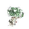
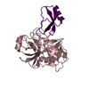
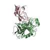
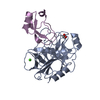
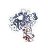
- PDB-2kai: REFINED 2.5 ANGSTROMS X-RAY CRYSTAL STRUCTURE OF THE COMPLEX FORM... -
+ Open data

- Basic information
| Entry | Database: PDB / ID: 2kai | ||||||
|---|---|---|---|---|---|---|---|
| Title | REFINED 2.5 ANGSTROMS X-RAY CRYSTAL STRUCTURE OF THE COMPLEX FORMED BY PORCINE KALLIKREIN A AND THE BOVINE PANCREATIC TRYPSIN INHIBITOR. CRYSTALLIZATION, PATTERSON SEARCH, STRUCTURE DETERMINATION, REFINEMENT, STRUCTURE AND COMPARISON WITH ITS COMPONENTS AND WITH THE BOVINE TRYPSIN-PANCREATIC TRYPSIN INHIBITOR COMPLEX | ||||||
 Components Components |
| ||||||
 Keywords Keywords | COMPLEX (PROTEINASE-INHIBITOR) | ||||||
| Function / homology |  Function and homology information Function and homology informationtissue kallikrein / regulation of systemic arterial blood pressure / sulfate binding / negative regulation of platelet aggregation / zymogen binding / potassium channel inhibitor activity / zymogen activation / molecular function inhibitor activity / negative regulation of thrombin-activated receptor signaling pathway / serine protease inhibitor complex ...tissue kallikrein / regulation of systemic arterial blood pressure / sulfate binding / negative regulation of platelet aggregation / zymogen binding / potassium channel inhibitor activity / zymogen activation / molecular function inhibitor activity / negative regulation of thrombin-activated receptor signaling pathway / serine protease inhibitor complex / secretory granule / serine-type endopeptidase inhibitor activity / protease binding / serine-type endopeptidase activity / calcium ion binding / : Similarity search - Function | ||||||
| Biological species |  | ||||||
| Method |  X-RAY DIFFRACTION / Resolution: 2.5 Å X-RAY DIFFRACTION / Resolution: 2.5 Å | ||||||
 Authors Authors | Bode, W. / Chen, Z. | ||||||
 Citation Citation | Journal: J.Mol.Biol. / Year: 1983 Title: Refined 2.5 A X-ray crystal structure of the complex formed by porcine kallikrein A and the bovine pancreatic trypsin inhibitor. Crystallization, Patterson search, structure determination, ...Title: Refined 2.5 A X-ray crystal structure of the complex formed by porcine kallikrein A and the bovine pancreatic trypsin inhibitor. Crystallization, Patterson search, structure determination, refinement, structure and comparison with its components and with the bovine trypsin-pancreatic trypsin inhibitor complex Authors: Chen, Z. / Bode, W. #1: Journal: J.Mol.Biol. / Year: 1983Title: Refined 2 Angstroms X-Ray Crystal Structure of Porcine Pancreatic Kallikrein A, a Specific Trypsin-Like Serine Proteinase. Crystallization, Structure Determination,Crystallographic ...Title: Refined 2 Angstroms X-Ray Crystal Structure of Porcine Pancreatic Kallikrein A, a Specific Trypsin-Like Serine Proteinase. Crystallization, Structure Determination,Crystallographic Refinement,Structure and its Comparison with Bovine Trypsin Authors: Bode, W. / Chen, Z. / Bartels, K. / Kutzbach, C. / Schmidt-Kastner, G. / Bartunik, H. | ||||||
| History |
|
- Structure visualization
| Structure viewer | Molecule: MolmilJmol/JSmol |
|---|
- Downloads & links
-Download
| PDBx/mmCIF format | 2kai.cif.gz | 65 KB | Display | PDBx/mmCIF format |
|---|---|---|---|---|
| PDB format | pdb2kai.ent.gz | 48.2 KB | Display | PDB format |
| PDBx/mmJSON format | 2kai.json.gz | Tree view | PDBx/mmJSON format | |
| Others |  Other downloads Other downloads |
-Validation report
| Arichive directory | https://data.pdbj.org/pub/pdb/validation_reports/ka/2kaiftp://data.pdbj.org/pub/pdb/validation_reports/ka/2kai | HTTPS FTP |
|---|
-Related structure data
| Similar structure data |
|---|


-Links
 PDBj
PDBj


- Assembly
| Deposited unit | 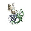
| ||||||||
|---|---|---|---|---|---|---|---|---|---|
| 1 |
| ||||||||
| Unit cell |
| ||||||||
| Atom site foot note | 1: RESIDUES PRO B 147, PRO B 219, PRO B 246 ARE CIS-PROLINES. ALSO SEE REMARK 5 IF OCCUPANCY IS 0.0. 2: SEE REMARK 5. / 3: SEE REMARK 6. |
-Components
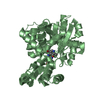
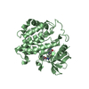
| #1: Protein | Mass: 9119.133 Da / Num. of mol.: 1 Source method: isolated from a genetically manipulated source Source: (gene. exp.) |
|---|---|
| #2: Protein | Mass: 16511.477 Da / Num. of mol.: 1 Source method: isolated from a genetically manipulated source Source: (gene. exp.) |
| #3: Protein | Mass: 6527.568 Da / Num. of mol.: 1 Source method: isolated from a genetically manipulated source Source: (gene. exp.) |
| #4: Water | ChemComp-HOH /  Mass: 18.015 Da / Num. of mol.: 10 / Source method: isolated from a natural source / Formula: H2O Mass: 18.015 Da / Num. of mol.: 10 / Source method: isolated from a natural source / Formula: H2O |
| Has protein modification | Y |
-Experimental details
-Experiment
| Experiment | Method: X-RAY DIFFRACTION |
|---|
- Sample preparation
| Crystal | Density Matthews: 4.76 Å3/Da / Density % sol: 74.15 % | |||||||||||||||
|---|---|---|---|---|---|---|---|---|---|---|---|---|---|---|---|---|
| Crystal grow | *PLUS Temperature: 20 ℃ / Method: vapor diffusion, hanging drop / PH range low: 4.25 / PH range high: 3.75 | |||||||||||||||
| Components of the solutions | *PLUS
|
-Data collection
| Radiation | Scattering type: x-ray |
|---|---|
| Radiation wavelength | Relative weight: 1 |
| Reflection | *PLUS Highest resolution: 2.5 Å / Num. obs: 16780 / Observed criterion σ(I): 2 / Num. measured all: 60000 / Rmerge(I) obs: 0.104 |
- Processing
| Software | Name: EREF / Classification: refinement | ||||||||||||
|---|---|---|---|---|---|---|---|---|---|---|---|---|---|
| Refinement | Rfactor Rwork: 0.224 / Highest resolution: 2.5 Å Details: AN OCCUPANCY OF 0.0 INDICATES THAT NO SIGNIFICANT ELECTRON DENSITY WAS FOUND IN THE FINAL FOURIER MAP AND THAT THE COORDINATES WERE GENERATED USING STEREOCHEMICAL CRITERIA. NO COORDINATES ...Details: AN OCCUPANCY OF 0.0 INDICATES THAT NO SIGNIFICANT ELECTRON DENSITY WAS FOUND IN THE FINAL FOURIER MAP AND THAT THE COORDINATES WERE GENERATED USING STEREOCHEMICAL CRITERIA. NO COORDINATES ARE INCLUDED FOR RESIDUE 1 OF PTI OR FOR MOST OF RESIDUE 58 OF PTI. IN ORDER NOT TO RESTRAIN THE APPROACH OF OG OF SER B 195 TO THE SUSCEPTIBLE INHIBITOR BOND, THE NORMAL CONSTRAINTS IMPOSED ON THE NON-BONDED INTERACTIONS BETWEEN THIS ATOM AND THE NEIGHBORING ATOMS OF THE INHIBITOR WERE REMOVED BY CONVERTING THIS OG INTO A NEW ATOM TYPE OI WITH NON-BONDING INTERACTION FORCES OF ZERO. THIS ATOM IS IDENTIFIED AS OG IN THE COORDINATES BELOW. | ||||||||||||
| Refinement step | Cycle: LAST / Highest resolution: 2.5 Å
| ||||||||||||
| Refinement | *PLUS Highest resolution: 2.5 Å / Rfactor obs: 0.224 | ||||||||||||
| Solvent computation | *PLUS | ||||||||||||
| Displacement parameters | *PLUS |
