Resolution: 2→41.96 Å / Cor.coef. Fo:Fc: 0.961 / Cor.coef. Fo:Fc free: 0.935 / SU B: 8.378 / SU ML: 0.119 / Cross valid method: THROUGHOUT / σ(F): 0 / σ(I): 2 / ESU R Free: 0.164 / Stereochemistry target values: MAXIMUM LIKELIHOOD / Details: HYDROGENS HAVE BEEN USED IN REFINEMENT
Rfactor
Num. reflection
% reflection
Selection details
Rfree
0.23095
1237
5.1 %
RANDOM
Rwork
0.16599
-
-
-
obs
0.16937
23165
99.09 %
-
all
-
24402
-
-
Solvent computation
Ion probe radii: 0.8 Å / Shrinkage radii: 0.8 Å / VDW probe radii: 1.2 Å / Solvent model: MASK
Displacement parameters
Biso mean: 24.314 Å2
Baniso -1
Baniso -2
Baniso -3
1-
-0.24 Å2
0 Å2
0 Å2
2-
-
1.95 Å2
0 Å2
3-
-
-
-1.71 Å2
Refine analyze
Luzzati coordinate error obs: 0.21 Å
Refinement step
Cycle: LAST / Resolution: 2→41.96 Å
Protein
Nucleic acid
Ligand
Solvent
Total
Num. atoms
2374
0
95
276
2745
Refine LS restraints
Refine-ID
Type
Dev ideal
Dev ideal target
Number
X-RAY DIFFRACTION
r_bond_refined_d
0.016
0.022
2584
X-RAY DIFFRACTION
r_bond_other_d
0.001
0.02
1783
X-RAY DIFFRACTION
r_angle_refined_deg
1.685
1.983
3489
X-RAY DIFFRACTION
r_angle_other_deg
0.903
3.002
4289
X-RAY DIFFRACTION
r_dihedral_angle_1_deg
6.134
5
310
X-RAY DIFFRACTION
r_dihedral_angle_2_deg
35.024
23.982
113
X-RAY DIFFRACTION
r_dihedral_angle_3_deg
13.322
15
435
X-RAY DIFFRACTION
r_dihedral_angle_4_deg
20.917
15
16
X-RAY DIFFRACTION
r_chiral_restr
0.093
0.2
381
X-RAY DIFFRACTION
r_gen_planes_refined
0.006
0.021
2797
X-RAY DIFFRACTION
r_gen_planes_other
0.001
0.02
505
LS refinement shell
Resolution: 2→2.052 Å / Total num. of bins used: 20
Rfactor
Num. reflection
% reflection
Rfree
0.334
68
-
Rwork
0.282
1588
-
obs
-
-
93.09 %
Refinement TLS params.
Method: refined / Refine-ID: X-RAY DIFFRACTION
ID
L11 (°2)
L12 (°2)
L13 (°2)
L22 (°2)
L23 (°2)
L33 (°2)
S11 (Å °)
S12 (Å °)
S13 (Å °)
S21 (Å °)
S22 (Å °)
S23 (Å °)
S31 (Å °)
S32 (Å °)
S33 (Å °)
T11 (Å2)
T12 (Å2)
T13 (Å2)
T22 (Å2)
T23 (Å2)
T33 (Å2)
Origin x (Å)
Origin y (Å)
Origin z (Å)
1
0.8451
-0.5098
-0.4208
1.1185
-0.4919
2.0173
0.0318
-0.0251
0.0397
0.0084
-0.0295
0.0442
-0.1666
-0.2262
-0.0023
0.0837
0.0341
0.0027
0.1172
0.0125
0.0585
1.8768
26.7266
23.7687
2
4.8017
-0.1935
-2.3236
1.0317
-0.2704
2.2862
-0.0075
0.1883
0.0859
0.0592
0.0597
-0.0252
-0.2964
-0.4026
-0.0522
0.112
0.0587
0.0194
0.1471
0.009
0.0231
3.4921
28.3995
25.5476
3
0.2451
-0.0439
-0.2347
0.1847
0.1428
1.2353
0.0145
0.0218
0.0505
0.0318
0.0156
0.0189
-0.1698
-0.0173
-0.0301
0.0566
0.0017
0.003
0.0643
0.0169
0.042
16.5387
25.8026
18.3211
4
0.2864
0.0051
-0.0578
0.1523
-0.1065
0.6709
0.0045
0.0372
-0.0314
0.0145
0.0051
0.017
0.0799
0.095
-0.0096
0.0394
0.0181
0.0049
0.0608
0.001
0.0453
19.2585
12.1463
14.93
5
0.202
0.5359
0.1003
1.9139
-0.6727
1.8463
0.021
0.0491
-0.0257
0.0162
-0.0645
-0.0958
0.0756
0.4193
0.0434
0.0106
0.0355
-0.0004
0.1402
0.0079
0.0415
31.2711
15.476
16.9115
Refinement TLS group
ID
Refine-ID
Refine TLS-ID
Auth asym-ID
Auth seq-ID
1
X-RAY DIFFRACTION
1
A
202 - 257
2
X-RAY DIFFRACTION
2
A
258 - 276
3
X-RAY DIFFRACTION
3
A
277 - 334
4
X-RAY DIFFRACTION
4
A
335 - 474
5
X-RAY DIFFRACTION
5
A
475 - 499
+
About Yorodumi
-
News
-
Feb 9, 2022. New format data for meta-information of EMDB entries
New format data for meta-information of EMDB entries
Version 3 of the EMDB header file is now the official format.
The previous official version 1.9 will be removed from the archive.
In the structure databanks used in Yorodumi, some data are registered as the other names, "COVID-19 virus" and "2019-nCoV". Here are the details of the virus and the list of structure data.
Jan 31, 2019. EMDB accession codes are about to change! (news from PDBe EMDB page)
EMDB accession codes are about to change! (news from PDBe EMDB page)
The allocation of 4 digits for EMDB accession codes will soon come to an end. Whilst these codes will remain in use, new EMDB accession codes will include an additional digit and will expand incrementally as the available range of codes is exhausted. The current 4-digit format prefixed with “EMD-” (i.e. EMD-XXXX) will advance to a 5-digit format (i.e. EMD-XXXXX), and so on. It is currently estimated that the 4-digit codes will be depleted around Spring 2019, at which point the 5-digit format will come into force.
The EM Navigator/Yorodumi systems omit the EMD- prefix.
Related info.:Q: What is EMD? / ID/Accession-code notation in Yorodumi/EM Navigator
Yorodumi is a browser for structure data from EMDB, PDB, SASBDB, etc.
This page is also the successor to EM Navigator detail page, and also detail information page/front-end page for Omokage search.
The word "yorodu" (or yorozu) is an old Japanese word meaning "ten thousand". "mi" (miru) is to see.
Related info.:EMDB / PDB / SASBDB / Comparison of 3 databanks / Yorodumi Search / Aug 31, 2016. New EM Navigator & Yorodumi / Yorodumi Papers / Jmol/JSmol / Function and homology information / Changes in new EM Navigator and Yorodumi
 Movie
Movie Controller

 Yorodumi
 Open data
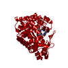
 Basic information
 Components
 Keywords
 Function and homology information
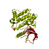
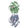
 Homo sapiens (human)
 X-RAY DIFFRACTION /
  Authors
 Citation
 Structure visualization
 Downloads & links
 Other downloads


 PDBj
PDBj
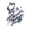
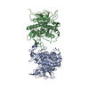
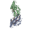
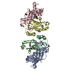
 Assembly


 Spodoptera frugiperda (fall armyworm)
 Mass: 94.971 Da / Num. of mol.: 2 / Source method: obtained synthetically / Formula: PO4
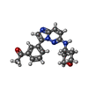
 Mass: 336.388 Da / Num. of mol.: 1 / Source method: obtained synthetically / Formula: C19H20N4O2
 Mass: 62.068 Da / Num. of mol.: 15 / Source method: obtained synthetically / Formula: C2H6O2
 Mass: 18.015 Da / Num. of mol.: 276 / Source method: isolated from a natural source / Formula: H2O
 Sample preparation
 / Beamline: I03 / Wavelength: 0.9763 Å
 Processing