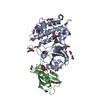
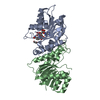
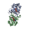
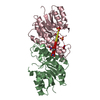
Entry Database : PDB / ID : 3h9rTitle Crystal structure of the kinase domain of type I activin receptor (ACVR1) in complex with FKBP12 and dorsomorphin Activin receptor type-1 Peptidyl-prolyl cis-trans isomerase FKBP1A Keywords / / / / / / / / / / / / / / / / / / / / Function / homology Function Domain/homology Component
/ / / / / / / / / / / / / / / / / / / / / / / / / / / / / / / / / / / / / / / / / / / / / / / / / / / / / / / / / / / / / / / / / / / / / / / / / / / / / / / / / / / / / / / / / / / / / / / / / / / / / / / / / / / / / / / / / / / / / / / / / / / / / / / / / / / / / / / / / / / / / / / / / / / / / / Biological species Homo sapiens (human)Method / / / Resolution : 2.35 Å Authors Chaikuad, A. / Alfano, I. / Shrestha, B. / Muniz, J.R.C. / Petrie, K. / Fedorov, O. / Phillips, C. / Bishop, S. / Mahajan, P. / Pike, A.C.W. ...Chaikuad, A. / Alfano, I. / Shrestha, B. / Muniz, J.R.C. / Petrie, K. / Fedorov, O. / Phillips, C. / Bishop, S. / Mahajan, P. / Pike, A.C.W. / von Delft, F. / Arrowsmith, C.H. / Edwards, A.M. / Weigelt, J. / Bountra, C. / Knapp, S. / Bullock, A. / Structural Genomics Consortium (SGC) Journal : J.Biol.Chem. / Year : 2012Title : Structure of the Bone Morphogenetic Protein Receptor ALK2 and Implications for Fibrodysplasia Ossificans Progressiva.Authors : Chaikuad, A. / Alfano, I. / Kerr, G. / Sanvitale, C.E. / Boergermann, J.H. / Triffitt, J.T. / von Delft, F. / Knapp, S. / Knaus, P. / Bullock, A.N. History Deposition Apr 30, 2009 Deposition site / Processing site Revision 1.0 Jun 2, 2009 Provider / Type Revision 1.1 Jul 13, 2011 Group / Version format complianceRevision 1.2 Nov 28, 2012 Group Revision 1.3 Sep 6, 2023 Group Data collection / Database references ... Data collection / Database references / Derived calculations / Refinement description Category chem_comp_atom / chem_comp_bond ... chem_comp_atom / chem_comp_bond / database_2 / pdbx_initial_refinement_model / struct_ref_seq_dif / struct_site Item _database_2.pdbx_DOI / _database_2.pdbx_database_accession ... _database_2.pdbx_DOI / _database_2.pdbx_database_accession / _struct_ref_seq_dif.details / _struct_site.pdbx_auth_asym_id / _struct_site.pdbx_auth_comp_id / _struct_site.pdbx_auth_seq_id
Show all Show less
 Movie
Movie Controller

 Yorodumi
 Open data
 Basic information
 Components
 Keywords
 Function and homology information
 Homo sapiens (human)
 X-RAY DIFFRACTION /
  Authors
 Citation
 Structure visualization
 Downloads & links
 Other downloads


 PDBj
PDBj


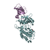
 Assembly
 Baculovirus


 Mass: 399.488 Da / Num. of mol.: 1 / Source method: obtained synthetically / Formula: C24H25N5O
 Mass: 96.063 Da / Num. of mol.: 5 / Source method: obtained synthetically / Formula: SO4
 Mass: 194.226 Da / Num. of mol.: 3 / Source method: obtained synthetically / Formula: C8H18O5 / Comment: precipitant*YM
 Sample preparation
 / Beamline: I02 / Wavelength: 0.905 Å
 Processing