 Movie
Movie Controller

[English] 日本語
 Yorodumi
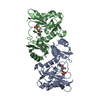
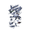
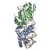
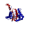
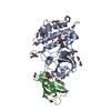
- PDB-1b6c: CRYSTAL STRUCTURE OF THE CYTOPLASMIC DOMAIN OF THE TYPE I TGF-BET... -
+ Open data

- Basic information
| Entry | Database: PDB / ID: 1b6c | ||||||
|---|---|---|---|---|---|---|---|
| Title | CRYSTAL STRUCTURE OF THE CYTOPLASMIC DOMAIN OF THE TYPE I TGF-BETA RECEPTOR IN COMPLEX WITH FKBP12 | ||||||
 Components Components |
| ||||||
 Keywords Keywords | COMPLEX (ISOMERASE/PROTEIN KINASE) / COMPLEX (ISOMERASE-PROTEIN KINASE) / RECEPTOR SERINE/THREONINE KINASE / COMPLEX (ISOMERASE-PROTEIN KINASE) complex | ||||||
| Function / homology |  Function and homology information Function and homology informationextracellular structure organization / epicardium morphogenesis / vascular endothelial cell proliferation / parathyroid gland development / regulation of cardiac muscle cell proliferation / transforming growth factor beta ligand-receptor complex / myofibroblast differentiation / positive regulation of epithelial to mesenchymal transition involved in endocardial cushion formation / TGFBR2 Kinase Domain Mutants in Cancer / transforming growth factor beta receptor activity ...extracellular structure organization / epicardium morphogenesis / vascular endothelial cell proliferation / parathyroid gland development / regulation of cardiac muscle cell proliferation / transforming growth factor beta ligand-receptor complex / myofibroblast differentiation / positive regulation of epithelial to mesenchymal transition involved in endocardial cushion formation / TGFBR2 Kinase Domain Mutants in Cancer / transforming growth factor beta receptor activity / trophoblast cell migration / SMAD2/3 Phosphorylation Motif Mutants in Cancer / TGFBR1 KD Mutants in Cancer / angiogenesis involved in coronary vascular morphogenesis / positive regulation of mesenchymal stem cell proliferation / ventricular compact myocardium morphogenesis / positive regulation of extracellular matrix assembly / positive regulation of tight junction disassembly / macrolide binding / cardiac epithelial to mesenchymal transition / transforming growth factor beta receptor activity, type I / mesenchymal cell differentiation / TGFBR3 regulates TGF-beta signaling / neuron fate commitment / positive regulation of vasculature development / activin receptor binding / activin receptor activity, type I / activin receptor complex / regulation of epithelial to mesenchymal transition / regulation of skeletal muscle contraction by regulation of release of sequestered calcium ion / cytoplasmic side of membrane / type II transforming growth factor beta receptor binding / pharyngeal system development / receptor protein serine/threonine kinase / activin binding / transmembrane receptor protein serine/threonine kinase activity / transforming growth factor beta receptor binding / TGFBR1 LBD Mutants in Cancer / primordial germ cell migration / embryonic cranial skeleton morphogenesis / coronary artery morphogenesis / filopodium assembly / activin receptor signaling pathway / type I transforming growth factor beta receptor binding / ventricular trabecula myocardium morphogenesis / negative regulation of activin receptor signaling pathway / signaling receptor inhibitor activity / heart trabecula formation / response to cholesterol / transforming growth factor beta binding / I-SMAD binding / collagen fibril organization / negative regulation of chondrocyte differentiation / lens development in camera-type eye / regulation of amyloid precursor protein catabolic process / terminal cisterna / ryanodine receptor complex / endothelial cell activation / anterior/posterior pattern specification / artery morphogenesis / positive regulation of filopodium assembly / skeletal system morphogenesis / 'de novo' protein folding / ventricular cardiac muscle tissue morphogenesis / ventricular septum morphogenesis / FK506 binding / negative regulation of endothelial cell proliferation / SMAD binding / roof of mouth development / TGF-beta receptor signaling activates SMADs / positive regulation of SMAD protein signal transduction / regulation of protein ubiquitination / mTORC1-mediated signalling / epithelial to mesenchymal transition / Calcineurin activates NFAT / bicellular tight junction / regulation of immune response / blastocyst development / cellular response to transforming growth factor beta stimulus / positive regulation of epithelial to mesenchymal transition / endothelial cell migration / heart morphogenesis / positive regulation of stress fiber assembly / supramolecular fiber organization / regulation of cardiac muscle contraction by regulation of the release of sequestered calcium ion / positive regulation of endothelial cell proliferation / sarcoplasmic reticulum membrane / transforming growth factor beta receptor signaling pathway / ciliary tip / negative regulation of cell migration / thymus development / T cell activation / peptidyl-serine phosphorylation / negative regulation of extrinsic apoptotic signaling pathway / Downregulation of TGF-beta receptor signaling / sarcoplasmic reticulum / post-embryonic development / peptidylprolyl isomerase / TGF-beta receptor signaling in EMT (epithelial to mesenchymal transition) / peptidyl-prolyl cis-trans isomerase activity Similarity search - Function | ||||||
| Biological species |  Homo sapiens (human) Homo sapiens (human) | ||||||
| Method |  X-RAY DIFFRACTION / SYNCHROTRON / MIR / Resolution: 2.6 Å X-RAY DIFFRACTION / SYNCHROTRON / MIR / Resolution: 2.6 Å | ||||||
 Authors Authors | Huse, M. / Chen, Y.-G. / Massague, J. / Kuriyan, J. | ||||||
 Citation Citation | Journal: Cell(Cambridge,Mass.) / Year: 1999 Title: Crystal structure of the cytoplasmic domain of the type I TGF beta receptor in complex with FKBP12. Authors: Huse, M. / Chen, Y.G. / Massague, J. / Kuriyan, J. | ||||||
| History |
|
- Structure visualization
| Structure viewer | Molecule: MolmilJmol/JSmol |
|---|
- Downloads & links
-Download
| PDBx/mmCIF format | 1b6c.cif.gz | 339.5 KB | Display | PDBx/mmCIF format |
|---|---|---|---|---|
| PDB format | pdb1b6c.ent.gz | 277.1 KB | Display | PDB format |
| PDBx/mmJSON format | 1b6c.json.gz | Tree view | PDBx/mmJSON format | |
| Others |  Other downloads Other downloads |
-Validation report
| Arichive directory | https://data.pdbj.org/pub/pdb/validation_reports/b6/1b6cftp://data.pdbj.org/pub/pdb/validation_reports/b6/1b6c | HTTPS FTP |
|---|
-Related structure data
| Similar structure data |
|---|


-Links
 PDBj
PDBj


- Assembly
| Deposited unit | 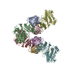
| ||||||||
|---|---|---|---|---|---|---|---|---|---|
| 1 | 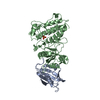
| ||||||||
| 2 | 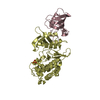
| ||||||||
| 3 | 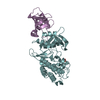
| ||||||||
| 4 | 
| ||||||||
| 5 | 
| ||||||||
| Unit cell |
| ||||||||
| Noncrystallographic symmetry (NCS) | NCS oper: (Code: given Matrix: (-0.999639, -0.026418, -0.004965), Vector: |
-Components
| #1: Protein | Mass: 11836.508 Da / Num. of mol.: 4 Source method: isolated from a genetically manipulated source Source: (gene. exp.) Homo sapiens (human) / Cell line: PLYS S / Plasmid: PFASTBAC / Cell line (production host): SF9 / Cellular location (production host): CYTOPLASM / Production host:   Spodoptera frugiperda (fall armyworm) / References: UniProt: P62942, peptidylprolyl isomerase Spodoptera frugiperda (fall armyworm) / References: UniProt: P62942, peptidylprolyl isomerase#2: Protein | Mass: 38920.641 Da / Num. of mol.: 4 / Fragment: CYTOPLASMIC PORTION Source method: isolated from a genetically manipulated source Source: (gene. exp.) Homo sapiens (human) / Cell line: PLYS S / Plasmid: PET23 / Cell line (production host): PLYS S / Cellular location (production host): CYTOPLASM / Production host:  #3: Chemical | ChemComp-SO4 /   Mass: 96.063 Da / Num. of mol.: 4 / Source method: obtained synthetically / Formula: SO4 Mass: 96.063 Da / Num. of mol.: 4 / Source method: obtained synthetically / Formula: SO4#4: Water | ChemComp-HOH / |  Mass: 18.015 Da / Num. of mol.: 88 / Source method: isolated from a natural source / Formula: H2O Mass: 18.015 Da / Num. of mol.: 88 / Source method: isolated from a natural source / Formula: H2O |
|---|
-Experimental details
-Experiment
| Experiment | Method: X-RAY DIFFRACTION / Number of used crystals: 6 |
|---|
- Sample preparation
| Crystal | Density Matthews: 2.6 Å3/Da / Density % sol: 52 % | ||||||||||||||||||||||||||||||||||||
|---|---|---|---|---|---|---|---|---|---|---|---|---|---|---|---|---|---|---|---|---|---|---|---|---|---|---|---|---|---|---|---|---|---|---|---|---|---|
| Crystal grow | pH: 8.5 / Details: pH 8.5 | ||||||||||||||||||||||||||||||||||||
| Crystal grow | *PLUS Temperature: 20 ℃ / Method: vapor diffusion | ||||||||||||||||||||||||||||||||||||
| Components of the solutions | *PLUS
|
-Data collection
| Diffraction | Mean temperature: 200 K |
|---|---|
| Diffraction source | Source: SYNCHROTRON / Site: NSLS  / Beamline: X25 / Wavelength: 1.5418 / Beamline: X25 / Wavelength: 1.5418 |
| Detector | Type: RIGAKU / Detector: IMAGE PLATE / Date: Feb 1, 1998 / Details: MIRRORS |
| Radiation | Monochromatic (M) / Laue (L): M / Scattering type: x-ray |
| Radiation wavelength | Wavelength: 1.5418 Å / Relative weight: 1 |
| Reflection | Resolution: 2.6→30 Å / Num. obs: 57740 / % possible obs: 98 % / Observed criterion σ(I): 0 / Redundancy: 3.5 % / Rsym value: 0.08 / Net I/σ(I): 18 |
| Reflection shell | Resolution: 2.6→2.69 Å / Redundancy: 3.5 % / Mean I/σ(I) obs: 6 / Rsym value: 0.24 / % possible all: 97.7 |
| Reflection | *PLUS Rmerge(I) obs: 0.08 |
| Reflection shell | *PLUS % possible obs: 97.7 % / Rmerge(I) obs: 0.243 |
- Processing
| Software |
| ||||||||||||||||||||||||||||||||||||||||||||||||||||||||||||
|---|---|---|---|---|---|---|---|---|---|---|---|---|---|---|---|---|---|---|---|---|---|---|---|---|---|---|---|---|---|---|---|---|---|---|---|---|---|---|---|---|---|---|---|---|---|---|---|---|---|---|---|---|---|---|---|---|---|---|---|---|---|
| Refinement | Method to determine structure: MIR / Resolution: 2.6→30 Å / Data cutoff high rms absF: 10000 / Isotropic thermal model: RESTRAINED / Cross valid method: THROUGHOUT / σ(F): 2
| ||||||||||||||||||||||||||||||||||||||||||||||||||||||||||||
| Solvent computation | Solvent model: FLAT | ||||||||||||||||||||||||||||||||||||||||||||||||||||||||||||
| Displacement parameters | Biso mean: 37.29 Å2
| ||||||||||||||||||||||||||||||||||||||||||||||||||||||||||||
| Refinement step | Cycle: LAST / Resolution: 2.6→30 Å
| ||||||||||||||||||||||||||||||||||||||||||||||||||||||||||||
| Refine LS restraints |
| ||||||||||||||||||||||||||||||||||||||||||||||||||||||||||||
| Refine LS restraints NCS | NCS model details: RESTRAINTS | ||||||||||||||||||||||||||||||||||||||||||||||||||||||||||||
| LS refinement shell | Resolution: 2.6→2.62 Å / Total num. of bins used: 50
| ||||||||||||||||||||||||||||||||||||||||||||||||||||||||||||
| Xplor file |
| ||||||||||||||||||||||||||||||||||||||||||||||||||||||||||||
| Software | *PLUS Name: CNS / Version: 0.3 / Classification: refinement | ||||||||||||||||||||||||||||||||||||||||||||||||||||||||||||
| Refinement | *PLUS | ||||||||||||||||||||||||||||||||||||||||||||||||||||||||||||
| Solvent computation | *PLUS | ||||||||||||||||||||||||||||||||||||||||||||||||||||||||||||
| Displacement parameters | *PLUS |
