 Movie
Movie Controller

[English] 日本語
 Yorodumi
- PDB-3tgv: Crystal structure of HutZ,the heme storsge protein from Vibrio ch... -
+ Open data

- Basic information
| Entry | Database: PDB / ID: 3tgv | ||||||
|---|---|---|---|---|---|---|---|
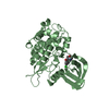
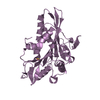
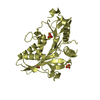
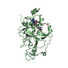
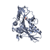
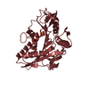
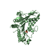
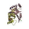
| Title | Crystal structure of HutZ,the heme storsge protein from Vibrio cholerae | ||||||
 Components Components | Heme-binding protein HutZ | ||||||
 Keywords Keywords | HEME BINDING PROTEIN | ||||||
| Function / homology |  Function and homology information Function and homology informationcoenzyme F420 binding / oxidoreductase activity, acting on the CH-CH group of donors / cytosol Similarity search - Function | ||||||
| Biological species |   Vibrio cholerae (bacteria) Vibrio cholerae (bacteria) | ||||||
| Method |  X-RAY DIFFRACTION / SYNCHROTRON / MOLECULAR REPLACEMENT / Resolution: 1.999 Å X-RAY DIFFRACTION / SYNCHROTRON / MOLECULAR REPLACEMENT / Resolution: 1.999 Å | ||||||
 Authors Authors | Liu, X. / Gong, J. / Wang, Z. / Du, Q. / Wei, T. / Zhu, D. / Huang, Y. / Xu, S. / Gu, L. | ||||||
 Citation Citation | Journal: To be Published Title: Crystal structure of HutZ,the heme storsge protein from Vibrio cholerae Authors: Liu, X. / Gong, J. / Wang, Z. / Du, Q. / Wei, T. / Zhu, D. / Huang, Y. / Xu, S. / Gu, L. | ||||||
| History |
|
- Structure visualization
| Structure viewer | Molecule: MolmilJmol/JSmol |
|---|
- Downloads & links
-Download
| PDBx/mmCIF format | 3tgv.cif.gz | 131.2 KB | Display | PDBx/mmCIF format |
|---|---|---|---|---|
| PDB format | pdb3tgv.ent.gz | 103 KB | Display | PDB format |
| PDBx/mmJSON format | 3tgv.json.gz | Tree view | PDBx/mmJSON format | |
| Others |  Other downloads Other downloads |
-Validation report
| Arichive directory | https://data.pdbj.org/pub/pdb/validation_reports/tg/3tgvftp://data.pdbj.org/pub/pdb/validation_reports/tg/3tgv | HTTPS FTP |
|---|
-Related structure data
| Related structure data |  1vl7S S: Starting model for refinement |
|---|---|
| Similar structure data |


-Links
 PDBj
PDBj- Assembly
| Deposited unit | 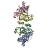
| ||||||||
|---|---|---|---|---|---|---|---|---|---|
| 1 | 
| ||||||||
| 2 | 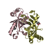
| ||||||||
| Unit cell |
|
-Components
| #1: Protein | Mass: 17320.729 Da / Num. of mol.: 4 / Fragment: UNP residues 13-150 / Mutation: H142Q Source method: isolated from a genetically manipulated source Source: (gene. exp.) Vibrio cholerae (bacteria) / Strain: O395 / Gene: hutZ / Production host: #2: Chemical | ChemComp-BEZ /   Mass: 122.121 Da / Num. of mol.: 4 / Source method: obtained synthetically / Formula: C7H6O2 Mass: 122.121 Da / Num. of mol.: 4 / Source method: obtained synthetically / Formula: C7H6O2#3: Water | ChemComp-HOH / |  Mass: 18.015 Da / Num. of mol.: 271 / Source method: isolated from a natural source / Formula: H2O Mass: 18.015 Da / Num. of mol.: 271 / Source method: isolated from a natural source / Formula: H2O |
|---|
-Experimental details
-Experiment
| Experiment | Method: X-RAY DIFFRACTION / Number of used crystals: 1 |
|---|
- Sample preparation
| Crystal | Density Matthews: 2.91 Å3/Da / Density % sol: 57.74 % |
|---|---|
| Crystal grow | Temperature: 293 K / Method: vapor diffusion, hanging drop / pH: 7.5 Details: 1.6M Na/K phosphate, 0.1M HEPES, pH 7.5, VAPOR DIFFUSION, HANGING DROP, temperature 293K |
-Data collection
| Diffraction | Mean temperature: 100 K |
|---|---|
| Diffraction source | Source: SYNCHROTRON / Site: SSRF  / Beamline: BL17U / Wavelength: 0.9793 Å / Beamline: BL17U / Wavelength: 0.9793 Å |
| Detector | Type: RAYONIX MX-225 / Detector: CCD / Date: Jun 10, 2009 |
| Radiation | Monochromator: Si(111) / Protocol: SINGLE WAVELENGTH / Monochromatic (M) / Laue (L): M / Scattering type: x-ray |
| Radiation wavelength | Wavelength: 0.9793 Å / Relative weight: 1 |
| Reflection | Resolution: 1.999→50 Å / Num. all: 51132 / Num. obs: 51132 / % possible obs: 95.5 % / Observed criterion σ(F): 0 / Observed criterion σ(I): 0 / Redundancy: 10 % / Rmerge(I) obs: 0.104 / Rsym value: 0.104 / Net I/σ(I): 24.7 |
| Reflection shell | Resolution: 2→2.07 Å / Redundancy: 5.9 % / Rmerge(I) obs: 0.474 / Mean I/σ(I) obs: 2.3 / Num. unique all: 3925 / Rsym value: 0.474 / % possible all: 73.9 |
- Processing
| Software |
| ||||||||||||||||||||||||||||||||||||||||||||||||||||||||||||||||||||||||||||||||||||||||||||||||||
|---|---|---|---|---|---|---|---|---|---|---|---|---|---|---|---|---|---|---|---|---|---|---|---|---|---|---|---|---|---|---|---|---|---|---|---|---|---|---|---|---|---|---|---|---|---|---|---|---|---|---|---|---|---|---|---|---|---|---|---|---|---|---|---|---|---|---|---|---|---|---|---|---|---|---|---|---|---|---|---|---|---|---|---|---|---|---|---|---|---|---|---|---|---|---|---|---|---|---|---|
| Refinement | Method to determine structure: MOLECULAR REPLACEMENT Starting model: PDB ENTRY 1VL7 Resolution: 1.999→35.805 Å / Occupancy max: 1 / Occupancy min: 1 / SU ML: 0.24 / σ(F): 0 / Phase error: 25.58 / Stereochemistry target values: ML
| ||||||||||||||||||||||||||||||||||||||||||||||||||||||||||||||||||||||||||||||||||||||||||||||||||
| Solvent computation | Shrinkage radii: 0.9 Å / VDW probe radii: 1.11 Å / Solvent model: FLAT BULK SOLVENT MODEL / Bsol: 40.416 Å2 / ksol: 0.386 e/Å3 | ||||||||||||||||||||||||||||||||||||||||||||||||||||||||||||||||||||||||||||||||||||||||||||||||||
| Displacement parameters | Biso max: 82.32 Å2 / Biso mean: 34.4221 Å2 / Biso min: 14.56 Å2
| ||||||||||||||||||||||||||||||||||||||||||||||||||||||||||||||||||||||||||||||||||||||||||||||||||
| Refinement step | Cycle: LAST / Resolution: 1.999→35.805 Å
| ||||||||||||||||||||||||||||||||||||||||||||||||||||||||||||||||||||||||||||||||||||||||||||||||||
| Refine LS restraints |
| ||||||||||||||||||||||||||||||||||||||||||||||||||||||||||||||||||||||||||||||||||||||||||||||||||
| LS refinement shell | Refine-ID: X-RAY DIFFRACTION / Total num. of bins used: 13
|
