 Movie
Movie Controller

[English] 日本語
 Yorodumi
- PDB-4m1z: Crystal structure of MycP1 with the N-terminal propeptide removed -
+ Open data

- Basic information
| Entry | Database: PDB / ID: 4m1z | ||||||
|---|---|---|---|---|---|---|---|
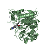
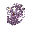
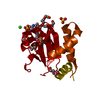
| Title | Crystal structure of MycP1 with the N-terminal propeptide removed | ||||||
 Components Components | (Membrane-anchored mycosin mycp1) x 2 | ||||||
 Keywords Keywords | HYDROLASE / subtilisin-like / propeptide-removed / serine protease / ESX-1 system / membrane-anchored | ||||||
| Function / homology |  Function and homology information Function and homology informationHydrolases; Acting on peptide bonds (peptidases); Serine endopeptidases / protein processing / serine-type endopeptidase activity / plasma membrane Similarity search - Function | ||||||
| Biological species |  Mycobacterium smegmatis (bacteria) Mycobacterium smegmatis (bacteria) | ||||||
| Method |  X-RAY DIFFRACTION / SYNCHROTRON / MOLECULAR REPLACEMENT / Resolution: 2.25 Å X-RAY DIFFRACTION / SYNCHROTRON / MOLECULAR REPLACEMENT / Resolution: 2.25 Å | ||||||
 Authors Authors | Sun, D.M. / He, Y. / Wang, C.L. / Zang, J.Y. / Tian, C.L. | ||||||
 Citation Citation | Journal: Protein Cell / Year: 2013 Title: The putative propeptide of MycP1 in mycobacterial type VII secretion system does not inhibit protease activity but improves protein stability. Authors: Sun, D.M. / Liu, Q. / He, Y. / Wang, C.L. / Wu, F.M. / Tian, C.L. / Zang, J.Y. | ||||||
| History |
|
- Structure visualization
| Structure viewer | Molecule: MolmilJmol/JSmol |
|---|
- Downloads & links
-Download
| PDBx/mmCIF format | 4m1z.cif.gz | 136.8 KB | Display | PDBx/mmCIF format |
|---|---|---|---|---|
| PDB format | pdb4m1z.ent.gz | 105.8 KB | Display | PDB format |
| PDBx/mmJSON format | 4m1z.json.gz | Tree view | PDBx/mmJSON format | |
| Others |  Other downloads Other downloads |
-Validation report
| Arichive directory | https://data.pdbj.org/pub/pdb/validation_reports/m1/4m1zftp://data.pdbj.org/pub/pdb/validation_reports/m1/4m1z | HTTPS FTP |
|---|
-Related structure data
| Related structure data | 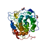 4kb5SC S: Starting model for refinement C: citing same article ( |
|---|---|
| Similar structure data |


-Links
 PDBj
PDBj

- Assembly
| Deposited unit | 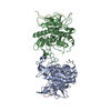
| ||||||||
|---|---|---|---|---|---|---|---|---|---|
| 1 | 
| ||||||||
| 2 | 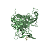
| ||||||||
| Unit cell |
|
-Components
| #1: Protein | Mass: 38626.934 Da / Num. of mol.: 2 / Fragment: UNP residues 63-422 Source method: isolated from a genetically manipulated source Source: (gene. exp.) Mycobacterium smegmatis (bacteria) / Strain: MC2 155 / Gene: MSMEG_0083, MSMEI_0081 / Plasmid: pET22b / Production host: #2: Protein/peptide | Mass: 2230.625 Da / Num. of mol.: 2 Source method: isolated from a genetically manipulated source Source: (gene. exp.) Mycobacterium smegmatis (bacteria) / Strain: MC2 155 / Gene: MSMEG_0083, MSMEI_0081 / Plasmid: pET22b / Production host: #3: Water | ChemComp-HOH / |  Mass: 18.015 Da / Num. of mol.: 291 / Source method: isolated from a natural source / Formula: H2O Mass: 18.015 Da / Num. of mol.: 291 / Source method: isolated from a natural source / Formula: H2OHas protein modification | Y | Sequence details | RESIDUES 63-402 IN THE CHAINS C AND D ARE DELETIONS. | |
|---|
-Experimental details
-Experiment
| Experiment | Method: X-RAY DIFFRACTION / Number of used crystals: 1 |
|---|
- Sample preparation
| Crystal | Density Matthews: 2.09 Å3/Da / Density % sol: 41.25 % |
|---|---|
| Crystal grow | Temperature: 283 K / Method: vapor diffusion, sitting drop / pH: 5.6 Details: 0.1M sodium citrate, 20% (v/v) 2-propanol, 20% (w/v) PEG 4000, pH 5.6, VAPOR DIFFUSION, SITTING DROP, temperature 283K |
-Data collection
| Diffraction | Mean temperature: 100 K |
|---|---|
| Diffraction source | Source: SYNCHROTRON / Site: SSRF  / Beamline: BL17U / Wavelength: 0.97906 Å / Beamline: BL17U / Wavelength: 0.97906 Å |
| Detector | Type: ADSC QUANTUM 315 / Detector: CCD / Date: Jun 18, 2013 |
| Radiation | Monochromator: Si 111 CHANNEL / Protocol: SINGLE WAVELENGTH / Monochromatic (M) / Laue (L): M / Scattering type: x-ray |
| Radiation wavelength | Wavelength: 0.97906 Å / Relative weight: 1 |
| Reflection | Resolution: 2.25→50 Å / Num. all: 33595 / Num. obs: 33595 / % possible obs: 99.5 % / Observed criterion σ(F): 0 / Observed criterion σ(I): -3 / Redundancy: 4 % / Biso Wilson estimate: 30.2 Å2 / Rmerge(I) obs: 0.102 / Rsym value: 0.102 / Net I/σ(I): 12.912 |
| Reflection shell | Resolution: 2.25→2.29 Å / Redundancy: 4.1 % / Rmerge(I) obs: 0.444 / Mean I/σ(I) obs: 2.845 / Rsym value: 0.444 / % possible all: 99.9 |
- Processing
| Software |
| |||||||||||||||||||||||||||||||||||||||||||||
|---|---|---|---|---|---|---|---|---|---|---|---|---|---|---|---|---|---|---|---|---|---|---|---|---|---|---|---|---|---|---|---|---|---|---|---|---|---|---|---|---|---|---|---|---|---|---|
| Refinement | Method to determine structure: MOLECULAR REPLACEMENT Starting model: PDB ENTRY 4KB5 Resolution: 2.25→37.28 Å / Cor.coef. Fo:Fc: 0.96 / Cor.coef. Fo:Fc free: 0.926 / SU B: 5.548 / SU ML: 0.139 / Cross valid method: THROUGHOUT / σ(F): 2 / ESU R: 0.297 / ESU R Free: 0.218 / Stereochemistry target values: MAXIMUM LIKELIHOOD / Details: HYDROGENS HAVE BEEN USED IF PRESENT IN THE INPUT
| |||||||||||||||||||||||||||||||||||||||||||||
| Solvent computation | Ion probe radii: 0.8 Å / Shrinkage radii: 0.8 Å / VDW probe radii: 1.2 Å / Solvent model: MASK | |||||||||||||||||||||||||||||||||||||||||||||
| Displacement parameters | Biso mean: 33.394 Å2
| |||||||||||||||||||||||||||||||||||||||||||||
| Refinement step | Cycle: LAST / Resolution: 2.25→37.28 Å
| |||||||||||||||||||||||||||||||||||||||||||||
| Refine LS restraints |
| |||||||||||||||||||||||||||||||||||||||||||||
| LS refinement shell | Resolution: 2.25→2.308 Å / Total num. of bins used: 20
|
