 Movie
Movie Controller


+ Open data

- Basic information
| Entry | Database: PDB / ID: 4kb5 | ||||||
|---|---|---|---|---|---|---|---|
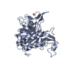
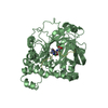
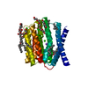
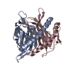
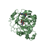
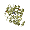
| Title | Crystal structure of MycP1 from Mycobacterium smegmatis | ||||||
 Components Components | Membrane-anchored mycosin mycp1 | ||||||
 Keywords Keywords | HYDROLASE / subtilisin-like serine protease / subtilase family / catalytic triad / autoinhibition / Serine protease | ||||||
| Function / homology |  Function and homology information Function and homology informationHydrolases; Acting on peptide bonds (peptidases); Serine endopeptidases / protein processing / serine-type endopeptidase activity / plasma membrane Similarity search - Function | ||||||
| Biological species |  Mycobacterium smegmatis (bacteria) Mycobacterium smegmatis (bacteria) | ||||||
| Method |  X-RAY DIFFRACTION / SYNCHROTRON / SAD / Resolution: 2.15 Å X-RAY DIFFRACTION / SYNCHROTRON / SAD / Resolution: 2.15 Å | ||||||
 Authors Authors | Sun, D.M. / He, Y. / Tian, C.L. | ||||||
 Citation Citation | Journal: Protein Cell / Year: 2013 Title: The putative propeptide of MycP1 in mycobacterial type VII secretion system does not inhibit protease activity but improves protein stability. Authors: Sun, D. / Liu, Q. / He, Y. / Wang, C. / Wu, F. / Tian, C. / Zang, J. | ||||||
| History |
|
- Structure visualization
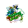
| Structure viewer | Molecule: MolmilJmol/JSmol |
|---|
- Downloads & links
-Download
| PDBx/mmCIF format | 4kb5.cif.gz | 90 KB | Display | PDBx/mmCIF format |
|---|---|---|---|---|
| PDB format | pdb4kb5.ent.gz | 66.9 KB | Display | PDB format |
| PDBx/mmJSON format | 4kb5.json.gz | Tree view | PDBx/mmJSON format | |
| Others |  Other downloads Other downloads |
-Validation report
| Arichive directory | https://data.pdbj.org/pub/pdb/validation_reports/kb/4kb5ftp://data.pdbj.org/pub/pdb/validation_reports/kb/4kb5 | HTTPS FTP |
|---|
-Related structure data


-Links
 PDBj
PDBj

- Assembly
| Deposited unit | 
| ||||||||
|---|---|---|---|---|---|---|---|---|---|
| 1 |
| ||||||||
| 2 | 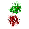
| ||||||||
| Unit cell |
|
-Components
| #1: Protein | Mass: 42502.203 Da / Num. of mol.: 1 / Fragment: UNP residues 24-422 Source method: isolated from a genetically manipulated source Source: (gene. exp.) Mycobacterium smegmatis (bacteria) / Strain: ATCC 700084 / mc(2)155 / Gene: MSMEG_0083, MSMEI_0081 / Production host: | ||||
|---|---|---|---|---|---|
| #2: Chemical | ChemComp-GOL /   Mass: 92.094 Da / Num. of mol.: 10 / Source method: obtained synthetically / Formula: C3H8O3 Mass: 92.094 Da / Num. of mol.: 10 / Source method: obtained synthetically / Formula: C3H8O3#3: Water | ChemComp-HOH / |  Mass: 18.015 Da / Num. of mol.: 177 / Source method: isolated from a natural source / Formula: H2O Mass: 18.015 Da / Num. of mol.: 177 / Source method: isolated from a natural source / Formula: H2OHas protein modification | Y | |
-Experimental details
-Experiment
| Experiment | Method: X-RAY DIFFRACTION / Number of used crystals: 1 |
|---|
- Sample preparation
| Crystal | Density Matthews: 4.12 Å3/Da / Density % sol: 70.11 % |
|---|---|
| Crystal grow | Temperature: 283 K / Method: vapor diffusion, sitting drop / pH: 5.5 Details: 1.6 M Magnesium sulfate, 0.1 M MES pH 5.5 with a protein concentration of 20 mg ml-1, VAPOR DIFFUSION, SITTING DROP, temperature 283K |
-Data collection
| Diffraction | Mean temperature: 100 K |
|---|---|
| Diffraction source | Source: SYNCHROTRON / Site: SSRF  / Beamline: BL17U / Wavelength: 0.97914 Å / Beamline: BL17U / Wavelength: 0.97914 Å |
| Detector | Type: MAR scanner 345 mm plate / Detector: IMAGE PLATE / Date: Jun 21, 2012 |
| Radiation | Monochromator: Si 111 CHANNEL / Protocol: SINGLE WAVELENGTH / Monochromatic (M) / Laue (L): M / Scattering type: x-ray |
| Radiation wavelength | Wavelength: 0.97914 Å / Relative weight: 1 |
| Reflection | Resolution: 2.15→77.89 Å / Num. all: 39312 / Num. obs: 39312 / % possible obs: 100 % / Observed criterion σ(F): 0 / Observed criterion σ(I): -3 / Redundancy: 7.6 % / Biso Wilson estimate: 34.6 Å2 / Rmerge(I) obs: 0.071 / Rsym value: 0.071 / Net I/σ(I): 25.9 |
| Reflection shell | Resolution: 2.15→2.19 Å / Redundancy: 7.7 % / Rmerge(I) obs: 0.507 / Mean I/σ(I) obs: 4.1 / Num. unique all: 1947 / Rsym value: 0.507 / % possible all: 99.8 |
- Processing
| Software |
| |||||||||||||||||||||||||||||||||||||||||||||
|---|---|---|---|---|---|---|---|---|---|---|---|---|---|---|---|---|---|---|---|---|---|---|---|---|---|---|---|---|---|---|---|---|---|---|---|---|---|---|---|---|---|---|---|---|---|---|
| Refinement | Method to determine structure: SAD / Resolution: 2.15→50 Å / Cor.coef. Fo:Fc: 0.959 / Cor.coef. Fo:Fc free: 0.948 / SU B: 3.691 / SU ML: 0.095 / Cross valid method: THROUGHOUT / σ(F): 2 / ESU R: 0.144 / ESU R Free: 0.134 / Stereochemistry target values: MAXIMUM LIKELIHOOD / Details: HYDROGENS HAVE BEEN USED IF PRESENT IN THE INPUT
| |||||||||||||||||||||||||||||||||||||||||||||
| Solvent computation | Ion probe radii: 0.8 Å / Shrinkage radii: 0.8 Å / VDW probe radii: 1.2 Å / Solvent model: MASK | |||||||||||||||||||||||||||||||||||||||||||||
| Displacement parameters | Biso mean: 39.261 Å2
| |||||||||||||||||||||||||||||||||||||||||||||
| Refinement step | Cycle: LAST / Resolution: 2.15→50 Å
| |||||||||||||||||||||||||||||||||||||||||||||
| Refine LS restraints |
| |||||||||||||||||||||||||||||||||||||||||||||
| LS refinement shell | Resolution: 2.151→2.207 Å / Total num. of bins used: 20
|
