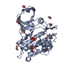
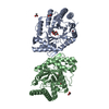
Entry Database : PDB / ID : 4dymTitle Crystal structure of the ACVR1 kinase domain in complex with the imidazo[1,2-b]pyridazine inhibitor K00135 Activin receptor type-1 Keywords / / / / / Function / homology Function Domain/homology Component
/ / / / / / / / / / / / / / / / / / / / / / / / / / / / / / / / / / / / / / / / / / / / / / / / / / / / / / / / / / / / / / / / / / / / / / / / / / / / / / / / / / / / / / / / / / / / / / / / / / / / / / / / Biological species Homo sapiens (human)Method / / / Resolution : 2.42 Å Authors Chaikuad, A. / Sanvitale, C. / Cooper, C. / Canning, P. / Mahajan, P. / Daga, N. / Petrie, K. / Alfano, I. / Gileadi, O. / Fedorov, O. ...Chaikuad, A. / Sanvitale, C. / Cooper, C. / Canning, P. / Mahajan, P. / Daga, N. / Petrie, K. / Alfano, I. / Gileadi, O. / Fedorov, O. / Krojer, T. / Filippakopoulos, P. / Muniz, J.R.C. / von Delft, F. / Weigelt, J. / Arrowsmith, C.H. / Edwards, A.M. / Bountra, C. / Bullock, A. / Structural Genomics Consortium (SGC) Journal : To be Published Title : Crystal structure of the ACVR1 kinase domain in complex with the imidazo[1,2-b]pyridazine inhibitor K00135Authors: Chaikuad, A. / Sanvitale, C. / Cooper, C. / Canning, P. / Mahajan, P. / Daga, N. / Petrie, K. / Alfano, I. / Gileadi, O. / Fedorov, O. / Krojer, T. / Filippakopoulos, P. / Muniz, J.R.C. / ... Authors : Chaikuad, A. / Sanvitale, C. / Cooper, C. / Canning, P. / Mahajan, P. / Daga, N. / Petrie, K. / Alfano, I. / Gileadi, O. / Fedorov, O. / Krojer, T. / Filippakopoulos, P. / Muniz, J.R.C. / von Delft, F. / Weigelt, J. / Arrowsmith, C.H. / Edwards, A.M. / Bountra, C. / Bullock, A. / Structural Genomics Consortium (SGC) History Deposition Feb 29, 2012 Deposition site / Processing site Revision 1.0 Mar 21, 2012 Provider / Type Revision 1.1 Sep 13, 2023 Group Data collection / Database references ... Data collection / Database references / Derived calculations / Refinement description Category chem_comp_atom / chem_comp_bond ... chem_comp_atom / chem_comp_bond / database_2 / pdbx_initial_refinement_model / struct_ref_seq_dif / struct_site Item _database_2.pdbx_DOI / _database_2.pdbx_database_accession ... _database_2.pdbx_DOI / _database_2.pdbx_database_accession / _struct_ref_seq_dif.details / _struct_site.pdbx_auth_asym_id / _struct_site.pdbx_auth_comp_id / _struct_site.pdbx_auth_seq_id
Show all Show less
 Movie
Movie Controller

 Yorodumi
 Open data
 Basic information
 Components
 Keywords
 Function and homology information
 Homo sapiens (human)
 X-RAY DIFFRACTION /
  Authors
 Citation
 Structure visualization
 Downloads & links
 Other downloads


 PDBj
PDBj
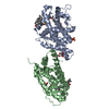
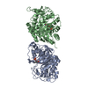
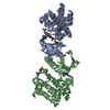
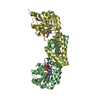
 Assembly
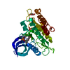


 Spodoptera frugiperda (fall armyworm)
 Mass: 96.063 Da / Num. of mol.: 2 / Source method: obtained synthetically / Formula: SO4
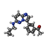
 Mass: 306.362 Da / Num. of mol.: 1 / Source method: obtained synthetically / Formula: C18H18N4O
 Mass: 92.094 Da / Num. of mol.: 4 / Source method: obtained synthetically / Formula: C3H8O3
 Mass: 18.015 Da / Num. of mol.: 124 / Source method: isolated from a natural source / Formula: H2O
 Sample preparation
 / Beamline: I02 / Wavelength: 0.9795 Å
 Processing