 Movie
Movie Controller


+ Open data

- Basic information
| Entry | Database: PDB / ID: 2iq1 | ||||||
|---|---|---|---|---|---|---|---|
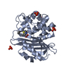
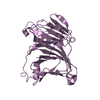
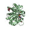
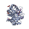
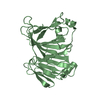
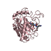
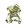
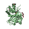
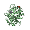
| Title | Crystal structure of human PPM1K | ||||||
 Components Components | Protein phosphatase 2C kappa, PPM1K | ||||||
 Keywords Keywords | HYDROLASE / protein phosphatase 2C kappa / structural genomics / PSI-2 / Protein Structure Initiative / New York SGX Research Center for Structural Genomics / NYSGXRC | ||||||
| Function / homology |  Function and homology information Function and homology information[3-methyl-2-oxobutanoate dehydrogenase (2-methylpropanoyl-transferring)]-phosphatase / [3-methyl-2-oxobutanoate dehydrogenase (lipoamide)]-phosphatase activity / regulation of intracellular signal transduction / H139Hfs13* PPM1K causes a mild variant of MSUD / branched-chain amino acid catabolic process / Branched-chain amino acid catabolism / regulation of mitochondrial membrane permeability involved in apoptotic process / protein-serine/threonine phosphatase / protein serine/threonine phosphatase activity / manganese ion binding ...[3-methyl-2-oxobutanoate dehydrogenase (2-methylpropanoyl-transferring)]-phosphatase / [3-methyl-2-oxobutanoate dehydrogenase (lipoamide)]-phosphatase activity / regulation of intracellular signal transduction / H139Hfs13* PPM1K causes a mild variant of MSUD / branched-chain amino acid catabolic process / Branched-chain amino acid catabolism / regulation of mitochondrial membrane permeability involved in apoptotic process / protein-serine/threonine phosphatase / protein serine/threonine phosphatase activity / manganese ion binding / mitochondrial matrix / mitochondrion Similarity search - Function | ||||||
| Biological species |  Homo sapiens (human) Homo sapiens (human) | ||||||
| Method |  X-RAY DIFFRACTION / SYNCHROTRON / SAD / Resolution: 2.25 Å X-RAY DIFFRACTION / SYNCHROTRON / SAD / Resolution: 2.25 Å | ||||||
 Authors Authors | Bonanno, J.B. / Freeman, J. / Russell, M. / Bain, K.T. / Adams, J. / Pelletier, L. / Wasserman, S. / Sauder, J.M. / Burley, S.K. / Almo, S.C. / New York SGX Research Center for Structural Genomics (NYSGXRC) | ||||||
 Citation Citation | Journal: J.STRUCT.FUNCT.GENOM. / Year: 2007 Title: Structural genomics of protein phosphatases Authors: Almo, S.C. / Bonanno, J.B. / Sauder, J.M. / Emtage, S. / Dilorenzo, T.P. / Malashkevich, V. / Wasserman, S.R. / Swaminathan, S. / Eswaramoorthy, S. / Agarwal, R. / Kumaran, D. / Madegowda, M. ...Authors: Almo, S.C. / Bonanno, J.B. / Sauder, J.M. / Emtage, S. / Dilorenzo, T.P. / Malashkevich, V. / Wasserman, S.R. / Swaminathan, S. / Eswaramoorthy, S. / Agarwal, R. / Kumaran, D. / Madegowda, M. / Ragumani, S. / Patskovsky, Y. / Alvarado, J. / Ramagopal, U.A. / Faber-Barata, J. / Chance, M.R. / Sali, A. / Fiser, A. / Zhang, Z.Y. / Lawrence, D.S. / Burley, S.K. | ||||||
| History |
|
- Structure visualization
| Structure viewer | Molecule: MolmilJmol/JSmol |
|---|
- Downloads & links
-Download
| PDBx/mmCIF format | 2iq1.cif.gz | 66.3 KB | Display | PDBx/mmCIF format |
|---|---|---|---|---|
| PDB format | pdb2iq1.ent.gz | 48.2 KB | Display | PDB format |
| PDBx/mmJSON format | 2iq1.json.gz | Tree view | PDBx/mmJSON format | |
| Others |  Other downloads Other downloads |
-Validation report
| Summary document | 2iq1_validation.pdf.gz | 427.9 KB | Display | wwPDB validaton report |
|---|---|---|---|---|
| Full document | 2iq1_full_validation.pdf.gz | 429.5 KB | Display | |
| Data in XML | 2iq1_validation.xml.gz | 12.7 KB | Display | |
| Data in CIF | 2iq1_validation.cif.gz | 17.5 KB | Display | |
| Arichive directory | https://data.pdbj.org/pub/pdb/validation_reports/iq/2iq1ftp://data.pdbj.org/pub/pdb/validation_reports/iq/2iq1 | HTTPS FTP |
-Related structure data
| Related structure data | 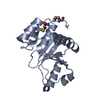 1rxdC 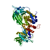 2fh7C 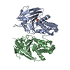 2g59C 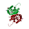 2hcmC  2hhlC 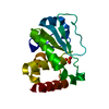 2hxpC  2hy3C 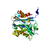 2i0oC 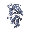 2i1yC 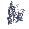 2i44C 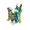 2irmC 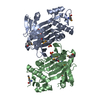 2isnC 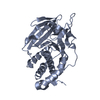 2nv5C 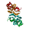 2oycC 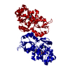 2p27C 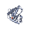 2p4uC 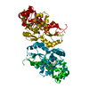 2p69C 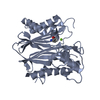 2p8eC 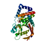 2pbnC 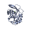 2q5eC 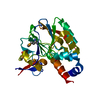 2qjcC 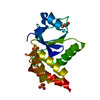 2r0bC C: citing same article ( |
|---|---|
| Similar structure data | |
| Other databases |

-Links
 PDBj
PDBj- Assembly
| Deposited unit | 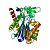
| ||||||||
|---|---|---|---|---|---|---|---|---|---|
| 1 |
| ||||||||
| Unit cell |
|
-Components
| #1: Protein | Mass: 30199.197 Da / Num. of mol.: 1 / Fragment: Residues 89-351 Source method: isolated from a genetically manipulated source Source: (gene. exp.) Homo sapiens (human) / Gene: DKFZp761G058 / Plasmid: modified pET26 / Species (production host): Escherichia coli / Production host:  |
|---|---|
| #2: Chemical | ChemComp-MG /   Mass: 24.305 Da / Num. of mol.: 1 / Source method: obtained synthetically / Formula: Mg Mass: 24.305 Da / Num. of mol.: 1 / Source method: obtained synthetically / Formula: Mg |
| #3: Water | ChemComp-HOH /  Mass: 18.015 Da / Num. of mol.: 126 / Source method: isolated from a natural source / Formula: H2O Mass: 18.015 Da / Num. of mol.: 126 / Source method: isolated from a natural source / Formula: H2O |
-Experimental details
-Experiment
| Experiment | Method: X-RAY DIFFRACTION / Number of used crystals: 1 |
|---|
- Sample preparation
| Crystal | Density Matthews: 2.64 Å3/Da / Density % sol: 53.46 % |
|---|---|
| Crystal grow | Temperature: 294 K / Method: vapor diffusion / pH: 8 Details: 100mM Hepes pH 8, 18% PEG 10K, VAPOR DIFFUSION, temperature 294K |
-Data collection
| Diffraction | Mean temperature: 77 K |
|---|---|
| Diffraction source | Source: SYNCHROTRON / Site: APS  / Beamline: 31-ID / Wavelength: 0.97958 Å / Beamline: 31-ID / Wavelength: 0.97958 Å |
| Detector | Type: MAR CCD 165 mm / Detector: CCD / Date: Oct 11, 2006 |
| Radiation | Monochromator: diamond / Protocol: SINGLE WAVELENGTH / Monochromatic (M) / Laue (L): M / Scattering type: x-ray |
| Radiation wavelength | Wavelength: 0.97958 Å / Relative weight: 1 |
| Reflection | Resolution: 2.25→51.674 Å / Num. all: 15773 / Num. obs: 15758 / % possible obs: 99.9 % / Observed criterion σ(F): 0 / Observed criterion σ(I): 0 / Redundancy: 7.8 % / Biso Wilson estimate: 38.4 Å2 / Rmerge(I) obs: 0.147 / Rsym value: 0.147 / Net I/σ(I): 15.7 |
| Reflection shell | Resolution: 2.25→2.37 Å / Redundancy: 8 % / Rmerge(I) obs: 0.848 / Mean I/σ(I) obs: 2.5 / Num. measured all: 17878 / Num. unique all: 2247 / Rsym value: 0.848 / % possible all: 100 |
-Phasing
| Phasing | Method: SAD |
|---|
- Processing
| Software |
| |||||||||||||||||||||||||||||||||||||||||||||||||||||||||||||||||||||||||||||||||||||||||||||||
|---|---|---|---|---|---|---|---|---|---|---|---|---|---|---|---|---|---|---|---|---|---|---|---|---|---|---|---|---|---|---|---|---|---|---|---|---|---|---|---|---|---|---|---|---|---|---|---|---|---|---|---|---|---|---|---|---|---|---|---|---|---|---|---|---|---|---|---|---|---|---|---|---|---|---|---|---|---|---|---|---|---|---|---|---|---|---|---|---|---|---|---|---|---|---|---|---|
| Refinement | Method to determine structure: SAD / Resolution: 2.25→20 Å / Cor.coef. Fo:Fc: 0.953 / Cor.coef. Fo:Fc free: 0.926 / SU B: 5.821 / SU ML: 0.148 / Cross valid method: THROUGHOUT / σ(F): 0 / σ(I): 0 / ESU R: 0.237 / ESU R Free: 0.206 / Stereochemistry target values: MAXIMUM LIKELIHOOD
| |||||||||||||||||||||||||||||||||||||||||||||||||||||||||||||||||||||||||||||||||||||||||||||||
| Solvent computation | Ion probe radii: 0.8 Å / Shrinkage radii: 0.8 Å / VDW probe radii: 1.4 Å / Solvent model: BABINET MODEL WITH MASK | |||||||||||||||||||||||||||||||||||||||||||||||||||||||||||||||||||||||||||||||||||||||||||||||
| Displacement parameters | Biso mean: 51.645 Å2
| |||||||||||||||||||||||||||||||||||||||||||||||||||||||||||||||||||||||||||||||||||||||||||||||
| Refinement step | Cycle: LAST / Resolution: 2.25→20 Å
| |||||||||||||||||||||||||||||||||||||||||||||||||||||||||||||||||||||||||||||||||||||||||||||||
| Refine LS restraints |
| |||||||||||||||||||||||||||||||||||||||||||||||||||||||||||||||||||||||||||||||||||||||||||||||
| LS refinement shell | Resolution: 2.25→2.308 Å / Total num. of bins used: 20
|
