 Movie
Movie Controller


+ Open data

- Basic information
| Entry | Database: PDB / ID: 2qjc | ||||||
|---|---|---|---|---|---|---|---|
| Title | Crystal structure of a putative diadenosine tetraphosphatase | ||||||
 Components Components | Diadenosine tetraphosphatase, putative | ||||||
 Keywords Keywords | HYDROLASE / 9095b / putative diadenosine tetraphosphatase / monomer / PSI-2 / Protein Structure Initiative / NYSGRC / Structural Genomics / New York SGX Research Center for Structural Genomics / NYSGXRC | ||||||
| Function / homology |  Function and homology information Function and homology informationpolyphosphate catabolic process / bis(5'-nucleosyl)-tetraphosphatase (symmetrical) / bis(5'-nucleosyl)-tetraphosphatase (symmetrical) activity / endopolyphosphatase activity / protein dephosphorylation / protein serine/threonine phosphatase activity / phosphatase activity / hydrolase activity / metal ion binding / cytoplasm Similarity search - Function | ||||||
| Biological species |  | ||||||
| Method |  X-RAY DIFFRACTION / SYNCHROTRON / SAD / Resolution: 2.05 Å X-RAY DIFFRACTION / SYNCHROTRON / SAD / Resolution: 2.05 Å | ||||||
 Authors Authors | Sugadev, R. / Burley, S.K. / Swaminathan, S. / New York SGX Research Center for Structural Genomics (NYSGXRC) | ||||||
 Citation Citation | Journal: J.Struct.Funct.Genom. / Year: 2007 Title: Structural genomics of protein phosphatases. Authors: Almo, S.C. / Bonanno, J.B. / Sauder, J.M. / Emtage, S. / Dilorenzo, T.P. / Malashkevich, V. / Wasserman, S.R. / Swaminathan, S. / Eswaramoorthy, S. / Agarwal, R. / Kumaran, D. / Madegowda, M. ...Authors: Almo, S.C. / Bonanno, J.B. / Sauder, J.M. / Emtage, S. / Dilorenzo, T.P. / Malashkevich, V. / Wasserman, S.R. / Swaminathan, S. / Eswaramoorthy, S. / Agarwal, R. / Kumaran, D. / Madegowda, M. / Ragumani, S. / Patskovsky, Y. / Alvarado, J. / Ramagopal, U.A. / Faber-Barata, J. / Chance, M.R. / Sali, A. / Fiser, A. / Zhang, Z.Y. / Lawrence, D.S. / Burley, S.K. | ||||||
| History |
|
- Structure visualization
| Structure viewer | Molecule: MolmilJmol/JSmol |
|---|
- Downloads & links
-Download
| PDBx/mmCIF format | 2qjc.cif.gz | 58.8 KB | Display | PDBx/mmCIF format |
|---|---|---|---|---|
| PDB format | pdb2qjc.ent.gz | 41.3 KB | Display | PDB format |
| PDBx/mmJSON format | 2qjc.json.gz | Tree view | PDBx/mmJSON format | |
| Others |  Other downloads Other downloads |
-Validation report
| Arichive directory | https://data.pdbj.org/pub/pdb/validation_reports/qj/2qjcftp://data.pdbj.org/pub/pdb/validation_reports/qj/2qjc | HTTPS FTP |
|---|
-Related structure data
| Related structure data | 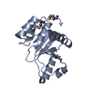 1rxdC 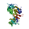 2fh7C 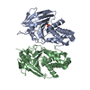 2g59C 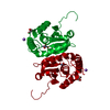 2hcmC  2hhlC  2hxpC  2hy3C  2i0oC 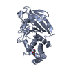 2i1yC 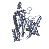 2i44C 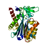 2iq1C 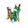 2irmC  2isnC  2nv5C  2oycC  2p27C  2p4uC  2p69C  2p8eC  2pbnC 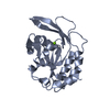 2q5eC 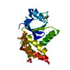 2r0bC C: citing same article ( |
|---|---|
| Similar structure data | |
| Other databases |


-Links
 PDBj
PDBj
- Assembly
| Deposited unit | 
| ||||||||
|---|---|---|---|---|---|---|---|---|---|
| 1 |
| ||||||||
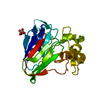
| Unit cell |
|
-Components
| #1: Protein | Mass: 28536.021 Da / Num. of mol.: 1 Source method: isolated from a genetically manipulated source Source: (gene. exp.)  References: UniProt: Q57U41, bis(5'-nucleosyl)-tetraphosphatase (symmetrical) | ||||||
|---|---|---|---|---|---|---|---|
| #2: Chemical |   Mass: 54.938 Da / Num. of mol.: 2 / Source method: obtained synthetically / Formula: Mn Mass: 54.938 Da / Num. of mol.: 2 / Source method: obtained synthetically / Formula: Mn#3: Chemical | ChemComp-PO4 / |   Mass: 94.971 Da / Num. of mol.: 1 / Source method: obtained synthetically / Formula: PO4 Mass: 94.971 Da / Num. of mol.: 1 / Source method: obtained synthetically / Formula: PO4#4: Water | ChemComp-HOH / |  Mass: 18.015 Da / Num. of mol.: 52 / Source method: isolated from a natural source / Formula: H2O Mass: 18.015 Da / Num. of mol.: 52 / Source method: isolated from a natural source / Formula: H2OHas protein modification | Y | |
-Experimental details
-Experiment
| Experiment | Method: X-RAY DIFFRACTION / Number of used crystals: 1 |
|---|
- Sample preparation
| Crystal | Density Matthews: 2.02 Å3/Da / Density % sol: 39.17 % |
|---|---|
| Crystal grow | Temperature: 298 K / Method: vapor diffusion, sitting drop / pH: 7 Details: 0.1M HEPES pH 7.0, 25% PEG 3350, VAPOR DIFFUSION, SITTING DROP, temperature 298K |
-Data collection
| Diffraction | Mean temperature: 100 K |
|---|---|
| Diffraction source | Source: SYNCHROTRON / Site: NSLS  / Beamline: X12C / Wavelength: 0.9795 Å / Beamline: X12C / Wavelength: 0.9795 Å |
| Detector | Type: ADSC QUANTUM 210 / Detector: CCD / Date: Jun 30, 2007 / Details: Mirrors |
| Radiation | Monochromator: Si(111) channel / Protocol: SINGLE WAVELENGTH / Monochromatic (M) / Laue (L): M / Scattering type: x-ray |
| Radiation wavelength | Wavelength: 0.9795 Å / Relative weight: 1 |
| Reflection | Resolution: 2.05→50 Å / Num. all: 15002 / Num. obs: 15002 / % possible obs: 99.4 % / Observed criterion σ(F): 0 / Observed criterion σ(I): 0 / Redundancy: 10.6 % / Biso Wilson estimate: 37.8 Å2 / Rmerge(I) obs: 0.07 / Net I/σ(I): 16 |
| Reflection shell | Resolution: 2.05→2.12 Å / Redundancy: 7.5 % / Rmerge(I) obs: 0.47 / Mean I/σ(I) obs: 1 / Num. unique all: 1428 / % possible all: 96.4 |
- Processing
| Software |
| ||||||||||||||||||||||||||||||||||||
|---|---|---|---|---|---|---|---|---|---|---|---|---|---|---|---|---|---|---|---|---|---|---|---|---|---|---|---|---|---|---|---|---|---|---|---|---|---|
| Refinement | Method to determine structure: SAD / Resolution: 2.05→49.4 Å / Rfactor Rfree error: 0.011 / Data cutoff high absF: 183986.4 / Data cutoff low absF: 0 / Isotropic thermal model: RESTRAINED / Cross valid method: THROUGHOUT / σ(F): 0 / Stereochemistry target values: Engh & Huber Details: Residues listed as missing in Remark 465 are due to lack of electron density. Residues with missing atoms listed in Remark 470 are due to lack of electron density for side chains and modeled as alanines.
| ||||||||||||||||||||||||||||||||||||
| Solvent computation | Solvent model: FLAT MODEL / Bsol: 55.5085 Å2 / ksol: 0.381149 e/Å3 | ||||||||||||||||||||||||||||||||||||
| Displacement parameters | Biso mean: 34.6 Å2
| ||||||||||||||||||||||||||||||||||||
| Refine analyze |
| ||||||||||||||||||||||||||||||||||||
| Refinement step | Cycle: LAST / Resolution: 2.05→49.4 Å
| ||||||||||||||||||||||||||||||||||||
| Refine LS restraints |
| ||||||||||||||||||||||||||||||||||||
| LS refinement shell | Resolution: 2.05→2.13 Å / Rfactor Rfree error: 0.051 / Total num. of bins used: 6
| ||||||||||||||||||||||||||||||||||||
| Xplor file |
|
