- PDB-2i4s: PDZ domain of EpsC from Vibrio cholerae, residues 204-305 -
+
Open data
ID or keywords:
Loading...
-
Basic information
Entry
Database: PDB / ID: 2i4s
Title
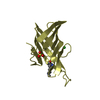
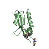
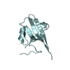
PDZ domain of EpsC from Vibrio cholerae, residues 204-305
Components
General secretion pathway protein C
Keywords
PROTEIN TRANSPORT / MEMBRANE PROTEIN / EpsC / GspC / PDZ domain / Type 2 Secretion System / General Secretion Pathway
Function / homology
Function and homology information
protein secretion by the type II secretion system / type II protein secretion system complex / plasma membrane Similarity search - Function
Type II secretion system protein GspC / Bacterial type II secretion system protein C signature. / Type II secretion system protein GspC, N-terminal / Type II secretion system protein C / PDZ domain / Pdz3 Domain / PDZ superfamily / Roll / Mainly Beta Similarity search - Domain/homology
Redundancy: 3.3 % / Av σ(I) over netI: 7 / Number: 50256 / Rmerge(I) obs: 0.093 / Χ2: 1.05 / D res high: 1.94 Å / D res low: 50 Å / Num. obs: 15285 / % possible obs: 97.7
Diffraction reflection shell
Highest resolution (Å)
Lowest resolution (Å)
% possible obs (%)
ID
Rmerge(I) obs
Chi squared
Redundancy
4.18
50
97.8
1
0.07
1.468
3
3.32
4.18
99.3
1
0.061
1.081
3.3
2.9
3.32
99.9
1
0.084
1.212
3.4
2.63
2.9
99.7
1
0.111
1.212
3.5
2.44
2.63
99.6
1
0.125
1.182
3.4
2.3
2.44
98.8
1
0.138
1.034
3.4
2.18
2.3
98.5
1
0.155
0.939
3.3
2.09
2.18
96.6
1
0.184
0.833
3.3
2.01
2.09
97
1
0.214
0.768
3.3
1.94
2.01
89.4
1
0.278
0.679
2.9
Reflection
Resolution: 1.94→50 Å / Num. obs: 15285 / % possible obs: 97.7 % / Redundancy: 3.3 % / Rmerge(I) obs: 0.093 / Χ2: 1.051 / Net I/σ(I): 7
Reflection shell
Resolution (Å)
Redundancy (%)
Rmerge(I) obs
Num. unique all
Χ2
% possible all
1.94-2.01
2.9
0.278
1370
0.679
89.4
2.01-2.09
3.3
0.214
1505
0.768
97
2.09-2.18
3.3
0.184
1481
0.833
96.6
2.18-2.3
3.3
0.155
1542
0.939
98.5
2.3-2.44
3.4
0.138
1511
1.034
98.8
2.44-2.63
3.4
0.125
1557
1.182
99.6
2.63-2.9
3.5
0.111
1548
1.212
99.7
2.9-3.32
3.4
0.084
1570
1.212
99.9
3.32-4.18
3.3
0.061
1571
1.081
99.3
4.18-50
3
0.07
1630
1.468
97.8
-
Processing
Software
Name
Version
Classification
NB
DENZO
datareduction
SCALEPACK
datascaling
REFMAC
refinement
PDB_EXTRACT
2
dataextraction
Refinement
Method to determine structure: SAD / Resolution: 1.92→50 Å / Cor.coef. Fo:Fc: 0.948 / Cor.coef. Fo:Fc free: 0.909 / SU B: 3.669 / SU ML: 0.109 / Cross valid method: THROUGHOUT / σ(F): 0 / ESU R: 0.18 / ESU R Free: 0.168 / Stereochemistry target values: MAXIMUM LIKELIHOOD / Details: HYDROGENS HAVE BEEN ADDED IN THE RIDING POSITIONS
Rfactor
Num. reflection
% reflection
Selection details
Rfree
0.236
759
5 %
RANDOM
Rwork
0.176
-
-
-
all
0.179
15282
-
-
obs
0.179
15282
96.94 %
-
Solvent computation
Ion probe radii: 0.8 Å / Shrinkage radii: 0.8 Å / VDW probe radii: 1.2 Å / Solvent model: MASK
Displacement parameters
Biso mean: 19.569 Å2
Baniso -1
Baniso -2
Baniso -3
1-
0.08 Å2
0 Å2
0.34 Å2
2-
-
-0.07 Å2
0 Å2
3-
-
-
0.37 Å2
Refinement step
Cycle: LAST / Resolution: 1.92→50 Å
Protein
Nucleic acid
Ligand
Solvent
Total
Num. atoms
1660
0
0
216
1876
Refine LS restraints
Refine-ID
Type
Dev ideal
Dev ideal target
Number
X-RAY DIFFRACTION
r_bond_refined_d
0.013
0.022
1682
X-RAY DIFFRACTION
r_angle_refined_deg
1.403
1.972
2270
X-RAY DIFFRACTION
r_dihedral_angle_1_deg
6.477
5
208
X-RAY DIFFRACTION
r_dihedral_angle_2_deg
31.606
25.556
90
X-RAY DIFFRACTION
r_dihedral_angle_3_deg
15.479
15
306
X-RAY DIFFRACTION
r_dihedral_angle_4_deg
20.766
15
12
X-RAY DIFFRACTION
r_chiral_restr
0.101
0.2
252
X-RAY DIFFRACTION
r_gen_planes_refined
0.005
0.02
1296
X-RAY DIFFRACTION
r_nbd_refined
0.254
0.3
776
X-RAY DIFFRACTION
r_nbtor_refined
0.311
0.5
1168
X-RAY DIFFRACTION
r_xyhbond_nbd_refined
0.212
0.5
280
X-RAY DIFFRACTION
r_symmetry_vdw_refined
0.232
0.3
62
X-RAY DIFFRACTION
r_symmetry_hbond_refined
0.199
0.5
38
X-RAY DIFFRACTION
r_mcbond_it
2.88
4
1071
X-RAY DIFFRACTION
r_mcangle_it
3.8
6
1686
X-RAY DIFFRACTION
r_scbond_it
5.234
6
667
X-RAY DIFFRACTION
r_scangle_it
6.312
8
584
LS refinement shell
Resolution: 1.924→1.974 Å / Total num. of bins used: 20
Rfactor
Num. reflection
% reflection
Rfree
0.378
42
-
Rwork
0.204
864
-
obs
-
906
77.24 %
+
About Yorodumi
-
News
-
Feb 9, 2022. New format data for meta-information of EMDB entries
New format data for meta-information of EMDB entries
Version 3 of the EMDB header file is now the official format.
The previous official version 1.9 will be removed from the archive.
In the structure databanks used in Yorodumi, some data are registered as the other names, "COVID-19 virus" and "2019-nCoV". Here are the details of the virus and the list of structure data.
Jan 31, 2019. EMDB accession codes are about to change! (news from PDBe EMDB page)
EMDB accession codes are about to change! (news from PDBe EMDB page)
The allocation of 4 digits for EMDB accession codes will soon come to an end. Whilst these codes will remain in use, new EMDB accession codes will include an additional digit and will expand incrementally as the available range of codes is exhausted. The current 4-digit format prefixed with “EMD-” (i.e. EMD-XXXX) will advance to a 5-digit format (i.e. EMD-XXXXX), and so on. It is currently estimated that the 4-digit codes will be depleted around Spring 2019, at which point the 5-digit format will come into force.
The EM Navigator/Yorodumi systems omit the EMD- prefix.
Related info.:Q: What is EMD? / ID/Accession-code notation in Yorodumi/EM Navigator
Yorodumi is a browser for structure data from EMDB, PDB, SASBDB, etc.
This page is also the successor to EM Navigator detail page, and also detail information page/front-end page for Omokage search.
The word "yorodu" (or yorozu) is an old Japanese word meaning "ten thousand". "mi" (miru) is to see.
Related info.:EMDB / PDB / SASBDB / Comparison of 3 databanks / Yorodumi Search / Aug 31, 2016. New EM Navigator & Yorodumi / Yorodumi Papers / Jmol/JSmol / Function and homology information / Changes in new EM Navigator and Yorodumi
 Movie
Movie Controller


 Open data
 Basic information
 Components
 Keywords
 Function and homology information
 Vibrio cholerae (bacteria)
 X-RAY DIFFRACTION /
  Authors
 Citation
 Structure visualization
 Downloads & links
 Other downloads


 PDBj
PDBj Assembly
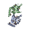

 Mass: 18.015 Da / Num. of mol.: 216 / Source method: isolated from a natural source / Formula: H2O
 Sample preparation
 / Beamline: 19-BM / Wavelength: 0.979124 Å
 Processing