 Movie
Movie Controller

[English] 日本語
 Yorodumi
- PDB-2sam: STRUCTURE OF THE PROTEASE FROM SIMIAN IMMUNODEFICIENCY VIRUS: COM... -
+ Open data

- Basic information
| Entry | Database: PDB / ID: 2sam | ||||||
|---|---|---|---|---|---|---|---|
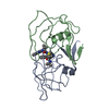
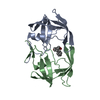
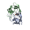
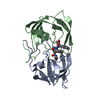
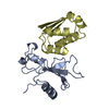
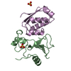
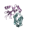
| Title | STRUCTURE OF THE PROTEASE FROM SIMIAN IMMUNODEFICIENCY VIRUS: COMPLEX WITH AN IRREVERSIBLE NON-PEPTIDE INHIBITOR | ||||||
 Components Components | SIV PROTEASE | ||||||
 Keywords Keywords | HYDROLASE(ACID PROTEASE) | ||||||
| Function / homology |  Function and homology information Function and homology informationexoribonuclease H activity / DNA integration / viral genome integration into host DNA / establishment of integrated proviral latency / RNA stem-loop binding / RNA-directed DNA polymerase activity / RNA-DNA hybrid ribonuclease activity / DNA recombination / aspartic-type endopeptidase activity / symbiont entry into host cell ...exoribonuclease H activity / DNA integration / viral genome integration into host DNA / establishment of integrated proviral latency / RNA stem-loop binding / RNA-directed DNA polymerase activity / RNA-DNA hybrid ribonuclease activity / DNA recombination / aspartic-type endopeptidase activity / symbiont entry into host cell / proteolysis / DNA binding / zinc ion binding Similarity search - Function | ||||||
| Biological species |  Simian immunodeficiency virus Simian immunodeficiency virus | ||||||
| Method |  X-RAY DIFFRACTION / Resolution: 2.4 Å X-RAY DIFFRACTION / Resolution: 2.4 Å | ||||||
 Authors Authors | Rose, R.B. / Rose, J.R. / Salto, R. / Craik, C.S. / Stroud, R.M. | ||||||
 Citation Citation | Journal: Biochemistry / Year: 1993 Title: Structure of the protease from simian immunodeficiency virus: complex with an irreversible nonpeptide inhibitor. Authors: Rose, R.B. / Rose, J.R. / Salto, R. / Craik, C.S. / Stroud, R.M. | ||||||
| History |
| ||||||
| Remark 700 | SHEET THE DIMER INTERFACE IS COMPOSED OF INTERDIGITATED N- AND C-TERMINI FROM BOTH SUBUNITS FORMING ...SHEET THE DIMER INTERFACE IS COMPOSED OF INTERDIGITATED N- AND C-TERMINI FROM BOTH SUBUNITS FORMING A FOUR-STRANDED ANTIPARALLEL BETA-SHEET. BECAUSE OF LIMITATIONS IMPOSED BY THE PROTEIN DATA BANK FORMAT IT IS NOT POSSIBLE TO PRESENT THIS SHEET ON SHEET RECORDS. INSTEAD THIS SHEET IS SPECIFIED IN THIS REMARK. STRANDS 1 AND 3 ARE FROM THE MOLECULE IN THIS ENTRY AND STRANDS 2 AND 4 ARE FROM THE SYMMETRY RELATED MOLECULE. I 4 PRO 1 THR 4 0 I 4 THR 96 PHE 99 -1 I 4 THR 96 PHE 99 -1 I 4 PRO 1 THR 4 -1 |
- Structure visualization
| Structure viewer | Molecule: MolmilJmol/JSmol |
|---|
- Downloads & links
-Download
| PDBx/mmCIF format | 2sam.cif.gz | 33.8 KB | Display | PDBx/mmCIF format |
|---|---|---|---|---|
| PDB format | pdb2sam.ent.gz | 21.7 KB | Display | PDB format |
| PDBx/mmJSON format | 2sam.json.gz | Tree view | PDBx/mmJSON format | |
| Others |  Other downloads Other downloads |
-Validation report
| Arichive directory | https://data.pdbj.org/pub/pdb/validation_reports/sa/2samftp://data.pdbj.org/pub/pdb/validation_reports/sa/2sam | HTTPS FTP |
|---|
-Related structure data
| Similar structure data |
|---|


-Links
 PDBj
PDBj- Assembly
| Deposited unit | 
| ||||||||
|---|---|---|---|---|---|---|---|---|---|
| 1 | 
| ||||||||
| Unit cell |
| ||||||||
| Atom site foot note | 1: THE EPN INHIBITOR IS COVALENTLY BOUND TO OD2 OF ASP 25. | ||||||||
| Components on special symmetry positions |
| ||||||||
| Details | THE HIV-1 PROTEASE IS A DIMER. IN THE CRYSTAL THE TWO MONOMERS ARE RELATED BY A CRYSTALLOGRAPHIC TWO-FOLD AXIS. TO GENERATE THE SYMMETRY RELATED MONOMER, THE FOLLOWING TRANSFORMATION MUST BE APPLIED TO THE COORDINATES PRESENTED IN THIS ENTRY MTRIX1 1 1.000000 0.000000 0.000000 0.000000 MTRIX2 1 0.000000 -1.000000 0.000000 0.000000 MTRIX3 1 0.000000 0.000000 -1.000000 0.000000 |
-Components
| #1: Protein | Mass: 10782.454 Da / Num. of mol.: 1 Source method: isolated from a genetically manipulated source Source: (gene. exp.) Simian immunodeficiency virus / Genus: Lentivirus / References: UniProt: Q88016, UniProt: Q5QGH9*PLUS |
|---|---|
| #2: Chemical | ChemComp-EPN /   Mass: 197.188 Da / Num. of mol.: 1 / Source method: obtained synthetically / Formula: C9H11NO4 Mass: 197.188 Da / Num. of mol.: 1 / Source method: obtained synthetically / Formula: C9H11NO4 |
| #3: Water | ChemComp-HOH /  Mass: 18.015 Da / Num. of mol.: 24 / Source method: isolated from a natural source / Formula: H2O Mass: 18.015 Da / Num. of mol.: 24 / Source method: isolated from a natural source / Formula: H2O |
| Has protein modification | Y |
| Nonpolymer details | THE OCCUPANCY OF THE LIGAND, EPNP, WAS REFINED TO 0.5. THE STOICHIOMETRY OF BINDING OF EPNP TO SIV ...THE OCCUPANCY OF THE LIGAND, EPNP, WAS REFINED TO 0.5. THE STOICHIOME |
| Sequence details | SEQUENCE ADVISORY NOTICE DIFFERENCE BETWEEN SWISS-PROT AND PDB SEQUENCE. SWISS-PROT ENTRY NAME: POL_ ...SEQUENCE ADVISORY NOTICE DIFFERENCE |
-Experimental details
-Experiment
| Experiment | Method: X-RAY DIFFRACTION |
|---|
- Sample preparation
| Crystal | Density Matthews: 2.25 Å3/Da / Density % sol: 45.28 % | ||||||||||||||||||||||||||||||||||||||||||
|---|---|---|---|---|---|---|---|---|---|---|---|---|---|---|---|---|---|---|---|---|---|---|---|---|---|---|---|---|---|---|---|---|---|---|---|---|---|---|---|---|---|---|---|
| Crystal grow | *PLUS pH: 6.5 / Method: vapor diffusion, hanging drop / Details: using macroseeding | ||||||||||||||||||||||||||||||||||||||||||
| Components of the solutions | *PLUS
|
-Data collection
| Reflection | *PLUS Highest resolution: 2.4 Å / Num. obs: 8824 / % possible obs: 92 % / Observed criterion σ(I): 1 / Num. measured all: 21880 / Rmerge(I) obs: 0.054 |
|---|---|
| Reflection shell | *PLUS Highest resolution: 2.25 Å / Lowest resolution: 2.5 Å / % possible obs: 77 % |
- Processing
| Software |
| ||||||||||||||||||||||||||||||||||||||||||||||||||||||||||||
|---|---|---|---|---|---|---|---|---|---|---|---|---|---|---|---|---|---|---|---|---|---|---|---|---|---|---|---|---|---|---|---|---|---|---|---|---|---|---|---|---|---|---|---|---|---|---|---|---|---|---|---|---|---|---|---|---|---|---|---|---|---|
| Refinement | Resolution: 2.4→40 Å / σ(F): 1 /
| ||||||||||||||||||||||||||||||||||||||||||||||||||||||||||||
| Refinement step | Cycle: LAST / Resolution: 2.4→40 Å
| ||||||||||||||||||||||||||||||||||||||||||||||||||||||||||||
| Refine LS restraints |
| ||||||||||||||||||||||||||||||||||||||||||||||||||||||||||||
| Software | *PLUS Name: X-PLOR / Classification: refinement | ||||||||||||||||||||||||||||||||||||||||||||||||||||||||||||
| Refinement | *PLUS Rfactor obs: 0.19 | ||||||||||||||||||||||||||||||||||||||||||||||||||||||||||||
| Solvent computation | *PLUS | ||||||||||||||||||||||||||||||||||||||||||||||||||||||||||||
| Displacement parameters | *PLUS | ||||||||||||||||||||||||||||||||||||||||||||||||||||||||||||
| Refine LS restraints | *PLUS Type: x_angle_d / Dev ideal: 3.2 |
