 Movie
Movie Controller


+ Open data

- Basic information
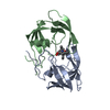
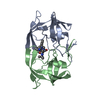
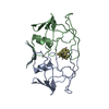
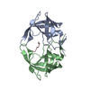
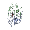
| Entry | Database: PDB / ID: 6upj | ||||||
|---|---|---|---|---|---|---|---|
| Title | HIV-2 PROTEASE/U99294 COMPLEX | ||||||
 Components Components | HIV-2 PROTEASE | ||||||
 Keywords Keywords | HYDROLASE / ACID PROTEASE / HIV-2 PROTEASE-INHIBITOR COMPLEX / PROTEIN-SUBSTRATE INTERACTION / ASPARTYL PROTEASE | ||||||
| Function / homology |  Function and homology information Function and homology informationHIV-2 retropepsin / retroviral ribonuclease H / exoribonuclease H / exoribonuclease H activity / DNA integration / viral genome integration into host DNA / establishment of integrated proviral latency / RNA-directed DNA polymerase / RNA stem-loop binding / viral penetration into host nucleus ...HIV-2 retropepsin / retroviral ribonuclease H / exoribonuclease H / exoribonuclease H activity / DNA integration / viral genome integration into host DNA / establishment of integrated proviral latency / RNA-directed DNA polymerase / RNA stem-loop binding / viral penetration into host nucleus / host multivesicular body / RNA-directed DNA polymerase activity / RNA-DNA hybrid ribonuclease activity / Transferases; Transferring phosphorus-containing groups; Nucleotidyltransferases / host cell / viral nucleocapsid / DNA recombination / DNA-directed DNA polymerase / aspartic-type endopeptidase activity / Hydrolases; Acting on ester bonds / DNA-directed DNA polymerase activity / symbiont-mediated suppression of host gene expression / viral translational frameshifting / symbiont entry into host cell / lipid binding / host cell plasma membrane / host cell nucleus / virion membrane / structural molecule activity / proteolysis / DNA binding / zinc ion binding Similarity search - Function | ||||||
| Biological species |  Human immunodeficiency virus 2 Human immunodeficiency virus 2 | ||||||
| Method |  X-RAY DIFFRACTION / MOLECULAR REPLACEMENT / Resolution: 2.34 Å X-RAY DIFFRACTION / MOLECULAR REPLACEMENT / Resolution: 2.34 Å | ||||||
 Authors Authors | Watenpaugh, K.D. / Mulichak, A.M. / Finzel, B.C. | ||||||
 Citation Citation | Journal: J.Med.Chem. / Year: 1995 Title: Use of medium-sized cycloalkyl rings to enhance secondary binding: discovery of a new class of human immunodeficiency virus (HIV) protease inhibitors. Authors: Romines, K.R. / Watenpaugh, K.D. / Tomich, P.K. / Howe, W.J. / Morris, J.K. / Lovasz, K.D. / Mulichak, A.M. / Finzel, B.C. / Lynn, J.C. / Horng, M.-M. / Schwende, F.J. / Ruwart, M.J. / Zipp, ...Authors: Romines, K.R. / Watenpaugh, K.D. / Tomich, P.K. / Howe, W.J. / Morris, J.K. / Lovasz, K.D. / Mulichak, A.M. / Finzel, B.C. / Lynn, J.C. / Horng, M.-M. / Schwende, F.J. / Ruwart, M.J. / Zipp, G.L. / Chong, K.-T. / Dolak, L.A. / Toth, L.N. / Howard, G.M. / Rush, B.D. / Wilkinson, K.F. / Possert, P.L. / Dalga, R.J. / Hinshaw, R.R. #1: Journal: J.Med.Chem. / Year: 1995 Title: Structure-based design of sulfonamide-substituted non-peptidic HIV protease inhibitors. Authors: Skulnick, H.I. / Johnson, P.D. / Howe, W.J. / Tomich, P.K. / Chong, K.-T. / Watenpaugh, K.D. / Janakiraman, M.N. / Dolak, L.A. / Mcgrath, J.P. / Lynn, J.C. / Horng, M.-M. / Hinshaw, R.R. / ...Authors: Skulnick, H.I. / Johnson, P.D. / Howe, W.J. / Tomich, P.K. / Chong, K.-T. / Watenpaugh, K.D. / Janakiraman, M.N. / Dolak, L.A. / Mcgrath, J.P. / Lynn, J.C. / Horng, M.-M. / Hinshaw, R.R. / Zipp, G.L. / Ruwart, M.J. / Schwende, F.J. / Zhong, W.-Z. / Padbury, G.E. / Dalga, R.J. / Shiou, L. / Possert, P.L. / Rush, B.D. / Wilkinson, K.F. / Howard, G.M. / Toth, L.N. / Williams, M.G. / Kakuk, T.J. / Cole, S.L. / Zaya, R.M. / Lovasz, K.D. / Morris, J.K. / Romines, K.R. / Thaisrivongs, S. / Aristoff, P.A. | ||||||
| History |
|
- Structure visualization
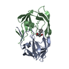
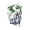
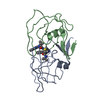
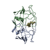
| Structure viewer | Molecule: MolmilJmol/JSmol |
|---|
- Downloads & links
-Download
| PDBx/mmCIF format | 6upj.cif.gz | 55.3 KB | Display | PDBx/mmCIF format |
|---|---|---|---|---|
| PDB format | pdb6upj.ent.gz | 38.8 KB | Display | PDB format |
| PDBx/mmJSON format | 6upj.json.gz | Tree view | PDBx/mmJSON format | |
| Others |  Other downloads Other downloads |
-Validation report
| Arichive directory | https://data.pdbj.org/pub/pdb/validation_reports/up/6upjftp://data.pdbj.org/pub/pdb/validation_reports/up/6upj | HTTPS FTP |
|---|
-Related structure data


-Links
 PDBj
PDBj
- Assembly
| Deposited unit | 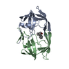
| ||||||||
|---|---|---|---|---|---|---|---|---|---|
| 1 |
| ||||||||
| Unit cell |
|
-Components
| #1: Protein | Mass: 10712.315 Da / Num. of mol.: 2 / Mutation: K57L Source method: isolated from a genetically manipulated source Source: (gene. exp.) Human immunodeficiency virus 2 / Genus: Lentivirus / Production host:  #2: Chemical | ChemComp-NIU / | 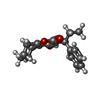  Mass: 298.376 Da / Num. of mol.: 1 / Source method: obtained synthetically / Formula: C19H22O3 Mass: 298.376 Da / Num. of mol.: 1 / Source method: obtained synthetically / Formula: C19H22O3#3: Water | ChemComp-HOH / |  Mass: 18.015 Da / Num. of mol.: 252 / Source method: isolated from a natural source / Formula: H2O Mass: 18.015 Da / Num. of mol.: 252 / Source method: isolated from a natural source / Formula: H2O |
|---|
-Experimental details
-Experiment
| Experiment | Method: X-RAY DIFFRACTION / Number of used crystals: 1 |
|---|
- Sample preparation
| Crystal | Density Matthews: 2.43 Å3/Da / Density % sol: 49.37 % | |||||||||||||||||||||||||
|---|---|---|---|---|---|---|---|---|---|---|---|---|---|---|---|---|---|---|---|---|---|---|---|---|---|---|
| Crystal grow | Method: vapor diffusion, hanging drop / pH: 7 Details: CRYSTALS WERE GROWN AT ROOM TEMPERATURE IN 4-7 UL HANGING DROPS OF EQUAL VOLUMES OF PROTEIN AT 8-10 MG/L AND WELL SOLUTION OF 30-35%(W/V) PEG 4000 IN 0.1 M HEPES AND PH RANGE 6.8 - 7.6., pH ...Details: CRYSTALS WERE GROWN AT ROOM TEMPERATURE IN 4-7 UL HANGING DROPS OF EQUAL VOLUMES OF PROTEIN AT 8-10 MG/L AND WELL SOLUTION OF 30-35%(W/V) PEG 4000 IN 0.1 M HEPES AND PH RANGE 6.8 - 7.6., pH 7.0, vapor diffusion - hanging drop PH range: 6.8-7.6 | |||||||||||||||||||||||||
| Crystal grow | *PLUS Method: vapor diffusion, hanging drop | |||||||||||||||||||||||||
| Components of the solutions | *PLUS
|
-Data collection
| Diffraction | Mean temperature: 298 K |
|---|---|
| Diffraction source | Source: ROTATING ANODE / Type: SIEMENS / Wavelength: 1.5418 |
| Detector | Type: SIEMENS / Detector: AREA DETECTOR / Date: Mar 9, 1993 |
| Radiation | Monochromator: GRAPHITE(002) / Monochromatic (M) / Laue (L): M / Scattering type: x-ray |
| Radiation wavelength | Wavelength: 1.5418 Å / Relative weight: 1 |
| Reflection | Resolution: 2.34→10 Å / Num. obs: 7557 / % possible obs: 81 % / Observed criterion σ(I): 0 / Redundancy: 3.7 % / Rmerge(I) obs: 0.071 / Net I/σ(I): 11.9 |
| Reflection shell | Resolution: 2.34→2.49 Å / Redundancy: 2 % / Rmerge(I) obs: 0.163 / Mean I/σ(I) obs: 4.37 / % possible all: 38 |
| Reflection | *PLUS Num. measured all: 27701 |
| Reflection shell | *PLUS % possible obs: 38 % |
- Processing
| Software |
| ||||||||||||||||||||||||||||||||||||||||||||||||||||||||||||
|---|---|---|---|---|---|---|---|---|---|---|---|---|---|---|---|---|---|---|---|---|---|---|---|---|---|---|---|---|---|---|---|---|---|---|---|---|---|---|---|---|---|---|---|---|---|---|---|---|---|---|---|---|---|---|---|---|---|---|---|---|---|
| Refinement | Method to determine structure: MOLECULAR REPLACEMENT Starting model: EARLIER STRUCTURE FROM SAME LABORATORY Resolution: 2.34→10 Å / σ(F): 2 /
| ||||||||||||||||||||||||||||||||||||||||||||||||||||||||||||
| Refinement step | Cycle: LAST / Resolution: 2.34→10 Å
| ||||||||||||||||||||||||||||||||||||||||||||||||||||||||||||
| Refine LS restraints |
| ||||||||||||||||||||||||||||||||||||||||||||||||||||||||||||
| Software | *PLUS Name: CEDAR / Classification: refinement | ||||||||||||||||||||||||||||||||||||||||||||||||||||||||||||
| Refinement | *PLUS Rfactor obs: 0.161 | ||||||||||||||||||||||||||||||||||||||||||||||||||||||||||||
| Solvent computation | *PLUS | ||||||||||||||||||||||||||||||||||||||||||||||||||||||||||||
| Displacement parameters | *PLUS |
